
|
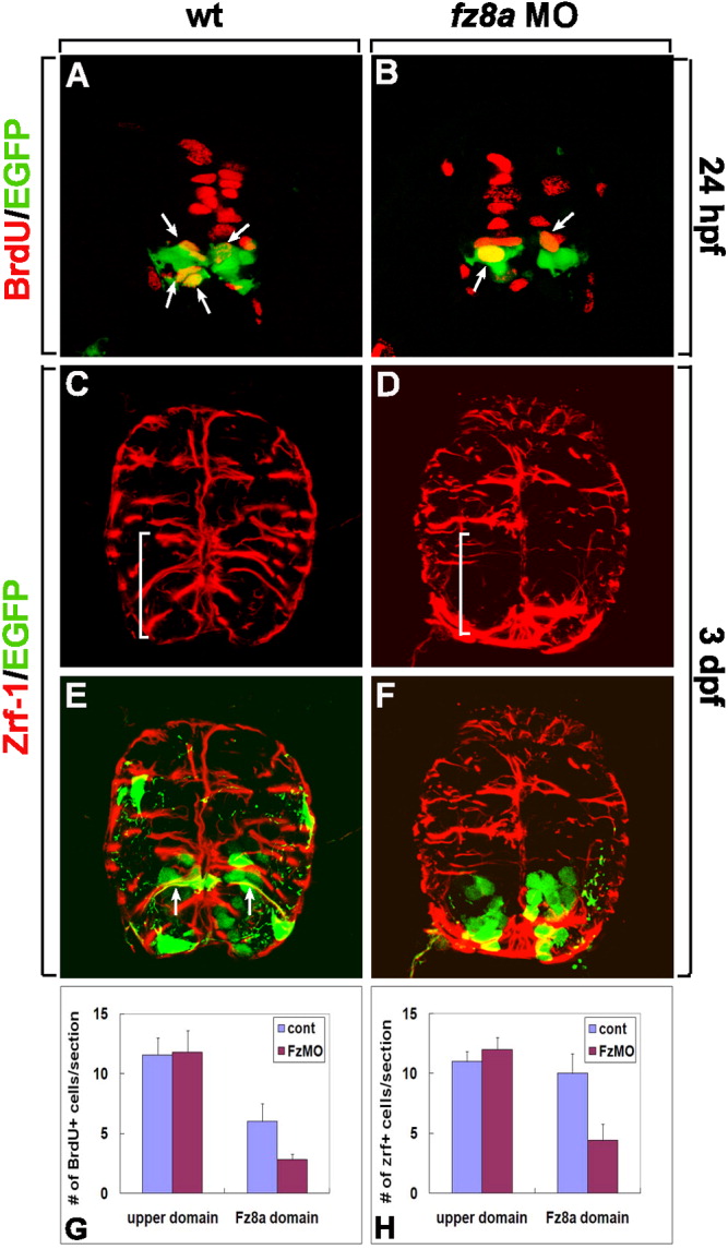
Fig. 4 fz8a function is required for the proliferation and spatial organization of radial glia in the ventral spinal cord. A-F: Transverse sections of spinal cords of Tg(olig2:egfp) embryos, dorsal up. A, B: Labeling of control (A) and fz8a MO-injected (B) embryos with an anti-BrdU antibody (red fluorescence) to detect proliferating cells. Arrows indicate olig2-expressing BrdU+ precursors. C-F: Combined anti-Zrf-1 (red fluorescence) and olig2:EGFP labeling of wt control (C, E, same section) and fz8a MO-injected (D, F, same section) embryos. fz8a MO-injected embryo shows disorganized radial glia in the ventral spinal cord (brackets). Arrows indicate processes of olig2+ radial glial cells labeled by olig2:EGFP and anti-Zrf-1 antibody staining (E). G: Quantification of BrdU+ proliferating cells at 24 hpf in control (cont) and fz8a MO-injected (FzMO) embryos. P < 0.0001 by Student's t-test for Fz8a domain. H: Quantification of the number of Zrf-1+ radial glial processes at 3 dpf in control (cont) and fz8a MO-injected (FzMO) embryos. Bars indicate the average number of cells per transverse section. Data were obtained from 10 sections from each of 8 control and 8 fz8a MO-injected embryos.