
|
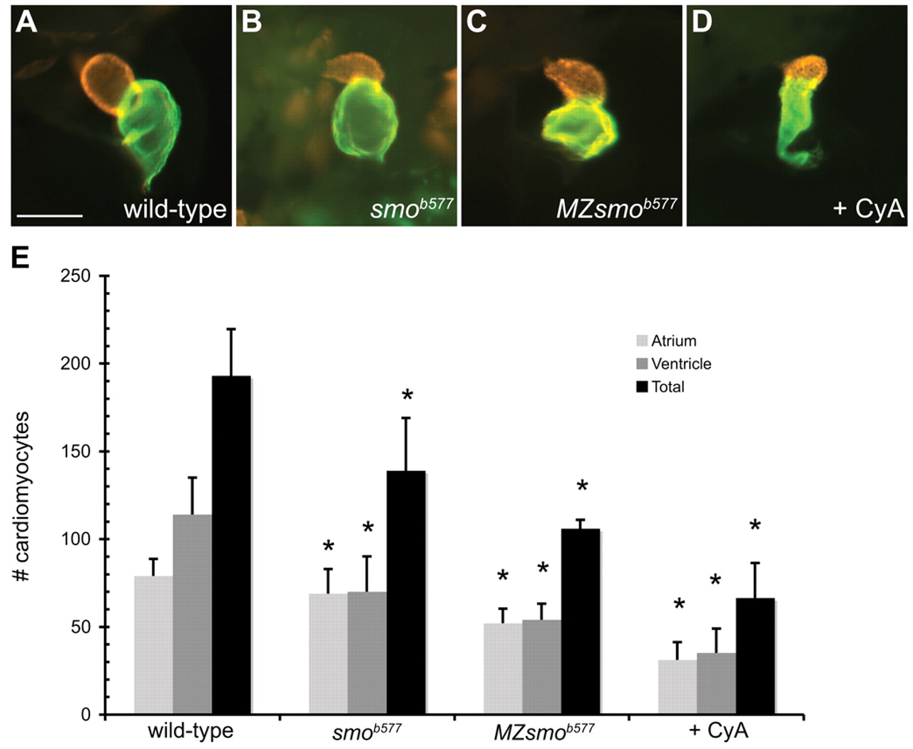
Fig. 1 Embryos with reduced Hedgehog signaling have small cardiac chambers. (A-D) Lateral views of zebrafish hearts stained with MF20 (red) and S46 (green) antibodies to visualize the ventricle and atrium at 48 hpf. MF20 marks the entire heart and S46 is atrium specific. In these superimposed images, the MF20+S46+ atrial tissue appears green and the MF20+S46- ventricle is red. Scale bar: 100 μm. (A) Wild-type heart. (B-D) Zygotic smob577 mutant, MZsmob577 mutant and CyA-treated hearts have small, misshapen ventricles and atria. (E) Quantification of cardiomyocyte number in wild-type, smob577, MZsmob577 and CyA-treated embryos at 52 hpf. See Materials and methods for cell counting technique. Values represent the mean cell number for each category (±s.d.). Asterisks indicate statistically significant differences from wild type (P<0.0001 for all except the smob577 atrium where P=0.01).