
|
Fig. 5
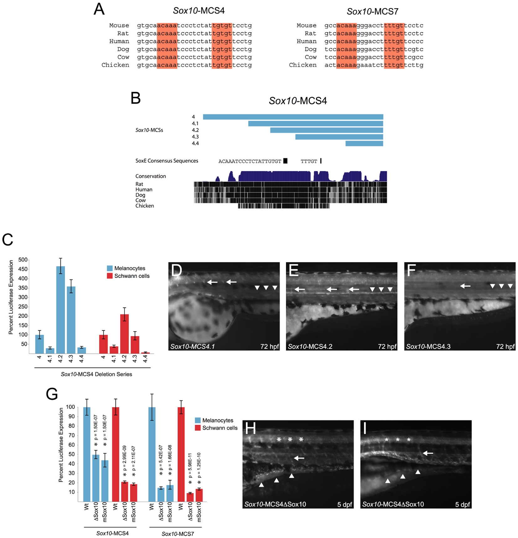
Analyses of a dimeric SoxE consensus sequence within Sox10-MCS4 and Sox10-MCS7.
A) Consensus SoxE family binding sites are oriented in a head-to-head fashion within Sox10-MCS4 and Sox10-MCS7. B) A deletion series across Sox10-MCS4 (pale blue bars) is depicted in the UCSC Genome Browser. The position and sequence of a monomeric and a dimeric SoxE consensus sequence is shown below the deletion series. C) In vitro enhancer activity for each fragment of the deletion series was tested individually in melan-a cells (blue bars) and S16 cells (red bars). The results for Sox10-MCS4 are included for cross-comparison of modular enhancer activity. Error bars, SD. D–F) in vivo enhancer activity was compared at 72 hpf for Sox10-MCS4.1 through Sox10-MCS4.3 in transgenic zebrafish embryos. Sox10-MCS4.4 failed to direct reporter expression in G0 embryos and was not raised for germline transmission. Sox10-MCS4.1 (D) directed reporter expression to Schwann cells (white arrows) and weak reporter expression to sympathetic ganglia (white arrowheads). Sox10-MCS4.2 (E) directed reporter expression in an opposite fashion, as signal appeared weaker in Schwann cells and more robust in sympathetic ganglia. Sox10-MCS4.3 (F) directed an extremely low level of reporter expression to these two neural crest–derived populations, and the arrow and arrowheads show the relative position of where the Schwann cells and sympathetic ganglia is normally positioned. G) Site-directed mutagenesis was used to delete and mutate the dimeric SoxE consensus sequence within Sox10-MCS4 and Sox10-MCS7. These mutagenized constructs were tested for in vitro enhancer activity in melanocytes (blue bars) and Schwann cells (red bars), and compared against their wild-type sequences. P-values are given above each tested construct and error bars indicate the standard deviation. H and I) Sox10-MCS4 with a deleted head-to-head SoxE family binding site was selected for transmission through the germline. Analysis of two different founder lines (H and I, respectively) revealed a decrease in signal in oligodendrocytes (asterisks) and scattered reporter expression in a subset of the ENS (white arrowheads). Schwann cells (white arrow), however, appear to be unaffected.