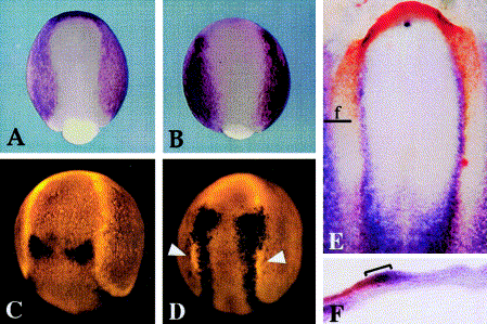
Fig. 3 Expression of Dlx and Msx genes in Xenopus, zebrafish, and chick. (A, B) Xenopus embryos at late gastrula stage showing expression of Dlx3 (A) and Msx1 (B). (C, D) Dorsal views of zebrafish embryos at late gastrula or early somitogenesis stages. (C) At bud stage, expression of dlx3 (red) is limited to the preplacodal domain and does not overlap with wnt8 in the hindbrain (black). By the 3-somite stage (D), expression of msxB (black) fully overlaps the dlx3 domain (red) in the preplacodal domain, except in the anterior head and in the lateral portion of the preotic domain (arrowheads). At this stage, msxB expression has started to shift medially into the neural plate. By the 10-somite stage, dlx3 and msxB totally separate into placodal and neural domains, respectively (not shown). (E, F) Chick embryo at stage 6 showing expression of Dlx5 (red) and Msx1 (blue). As seen in a wholemount specimen (E), Msx1 overlaps with Dlx5 except in the anterior head region. The plane of section for (F) is indicated. (F) A section confirms that Dlx5 and Msx1 overlap in the medial preplacodal domain (bracket). Images show dorsal views with anterior to the top (A–E) or a cross section with lateral to the left and dorsal to the top (F). With permission, (A) and (B) are reprinted from Feledy et al. (2001) and (E) and (F) are reprinted from [Streit 2002].
Reprinted from Developmental Biology, 261(2), Riley, B.B. and Phillips, B.T., Ringing in the new ear: resolution of cell interactions in otic development, 289-312, Copyright (2003) with permission from Elsevier. Full text @ Dev. Biol.

