
|
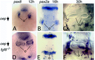
Fig. 2 Ear development in embryos deficient in oep and fgf8. (A–C) Wild-type embryos injected with oep-MO as previously described [Phillips et al 2001]. Small bilateral patches of pax8 at 12 h (A) or pax2a at 16 h (B) mark the developing otic placodes (o). By 30 h, otic vesicles form bilaterally but often elongate medially to touch at the midline. However, they are never observed to fuse (over 300 embryos examined). (D–F) ace/+ intercross progeny injected with oep-MO. About 25% of injected progeny showed dramatic changes in gene expression and otic vesicle morphology. These are inferred to be ace/ace(fgf8-/-) mutants. At 12 h, the otic domain of pax8 forms bilateral transverse bands that nearly touch at the midline (D) in 27% (12/44) of embryos. By 16 h, pax2a is expressed in a contiguous transverse stripe through the hindbrain (E) in 19% (9/48) of embryos. At 30 h, a single large otic vesicle forms at the midline and fully spans the width of the hindbrain (F) in 27% (15/55) of embryos. All images are dorsal views with anterior to the top. Abbreviation: o, otic tissue.
Reprinted from Developmental Biology, 261(2), Riley, B.B. and Phillips, B.T., Ringing in the new ear: resolution of cell interactions in otic development, 289-312, Copyright (2003) with permission from Elsevier. Full text @ Dev. Biol.