
|
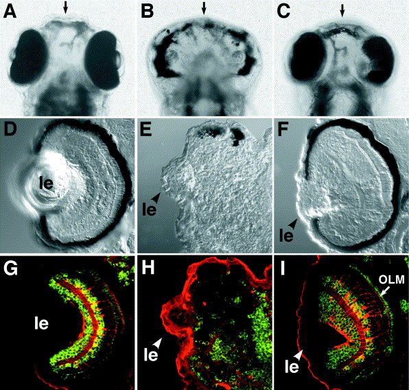
Fig. 3 Rescue of the glom117 phenotype by N-cadherin mRNA expression. Eye pigmentation and retinal architecture of the wild type (A, D, G), glom117 (B, E, H), and rescued glom117 embryos (C, F, I) at 4 dpf. (A–C) Ventral views of whole embryos. (D–F) Images of transverse sections through retinae. In (G–I), transverse sections of the retina were costained with anti-Hu (green, stains ganglion and amacrine cells), Zpr-1 (green, stains cone photoreceptor cells), and anti-carbonic anhydrase (red, stains Muller glia) antibodies. Note that the architecture of Muller glia and the outer limiting membrane (OLM, arrow) are restored in rescued animals. For rescued embryos, mutant genotype was confirmed by using a polymorphic marker linked to the N-cadherin locus. In (D–J), “le” indicates lens, dorsal is up. Arrows in (A–C) indicate the midline.
Reprinted from Developmental Biology, 259(1), Malicki, J., Jo, H., and Pujic, Z., Zebrafish N-cadherin, encoded by the glass onion locus, plays an essential role in retinal patterning, 95-108, Copyright (2003) with permission from Elsevier. Full text @ Dev. Biol.