
|
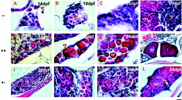
Fig. 5 Gonadal histology of TG(β-actin:EGFP) in --, ++, and +- groups juveniles. (A–D) Transverse sections showing germ cell development in -- group juveniles. Germ cells of 14- to 26-dpf-old juveniles in -- group are solely composed of gonocytes and surrounded by somatic cells. (E–H) Transverse sections showing germ cell development in ++ group juveniles. Germ cells of 14- to 26-dpf-old juveniles in ++ group juveniles are solely composed of early diplotene oocytes. (I–L) Horizontal sections showing germ cell development in +- group juveniles. Degenerating diplotene oocytes, gonocytes, and spermatogonia coexist in the gonads of 26-dpf-old juveniles in the +- group. Developmental stages are indicated in each panel. dpf, day postfertilization; ed, early diplotene oocytes; go, gonocytes; po, perinucleolar oocytes; sc, somatic cells; sg, spermatogonia; so, spermatocytes; sp, spermatids. Scale BAR = 25 μm in (E–I) and 10 μm in all other pictures.
Reprinted from Developmental Biology, 262, Hsiao, C.-D. and Tsai, H.-J., Transgenic zebrafish with fluorescent germ cell: a useful tool to visualize germ cell proliferation and juvenile hermaphroditism in vivo, 313-323, Copyright (2003) with permission from Elsevier. Full text @ Dev. Biol.