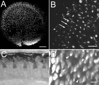
Fig. 5 Rod photoreceptor mosaic development in the larval retina. (A) Merged stack of low magnification confocal images of EGFP expression in the eye from a 10-dpf larval fish. The fluorescence appears scattered with regions of high density and obvious gaps in the distribution of rods. (B) At a higher magnification, the regular arrangement of parallel row of rods is indicated (arrows). (C) Histological section to demonstrate the regular spacing of the short single cones and the double cones in the larval retina. (D) Confocal image of EGFP fluorescence in larva 21 dpf. Note the regular arrangement of rows and the numerous telodendria extending from the rod terminals (arrowheads) Bars, 50 μm (A), 10 μm (B), 7 μm (C, D).
Reprinted from Developmental Biology, 258(2), Fadool, J.M., Development of a rod photoreceptor mosaic revealed in transgenic zebrafish, 277-290, Copyright (2003) with permission from Elsevier. Full text @ Dev. Biol.

