
Image
|
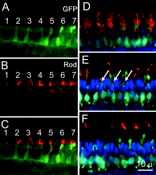
Figure Caption
Fig. 4 Rod differentiation at the retinal margin. (A–C) EGFP expression, immunolabeling for rhodopsin (Rod), and the merged image of an oblique section through the retinal margin reveal regularly spaced clusters of rods (numbered 1–7). (D–F) Merged images of immunolabeling (red) for rhodopsin (D), UV opsin (E), and red opsin (F) with EGFP expression (green) and DAPI nuclear staining (blue) of serial sections taken parallel to the retinal margin. Note the positions of the clustered rod outer segments at the positions of the immature UV cone outer segments (arrows).
Acknowledgments
This image is the copyrighted work of the attributed author or publisher, and
ZFIN has permission only to display this image to its users.
Additional permissions should be obtained from the applicable author or publisher of the image.
Reprinted from Developmental Biology, 258(2), Fadool, J.M., Development of a rod photoreceptor mosaic revealed in transgenic zebrafish, 277-290, Copyright (2003) with permission from Elsevier. Full text @ Dev. Biol.