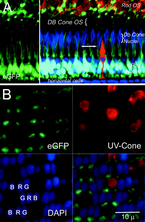
Fig. 3 Comparison of the rod mosaic to cone mosaic. (A) Transverse section through the adult retina demonstrating EGFP fluorescence in rod photoreceptors (left panel in green) and counterstaining with DAPI (blue) to reveal the tiering of the cone nuclei (left merged image). The rod cell bodies appear white due to double labeling for EGFP and DAPI. The rod outer segments are also immunolabeled for opsin. The position of the short single cone is diagrammatically represented and the plane of section in (B) is indicated by the arrow. (B) EGFP fluorescence of the rods, immunolabeling of the UV sensitive opsin, DAPI labeling of the cone nuclei in a tangential section of the adult retina and the merged image. Note the regular arrangement of the rod myoids around the outer segment of the UV-cone outer segments. (B, blue cone; G, green cone; R, red cone).
Reprinted from Developmental Biology, 258(2), Fadool, J.M., Development of a rod photoreceptor mosaic revealed in transgenic zebrafish, 277-290, Copyright (2003) with permission from Elsevier. Full text @ Dev. Biol.

