
|
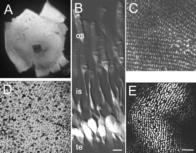
Fig. 2 Confocal analysis of EGFP expression of rod photoreceptors. Whole retinas maintained in saline were examined by confocal microscopy. (A) Low magnification image of the whole explant demonstrated robust expression of EGFP across the entire retina. (B) A higher magnification composite image of scans taken near the retinal margin. Cells with a characteristic rod morphology highlighted by small terminals (te), round soma, long thin inner segments (is), and rod-shaped outer segments (os) were observed. Images taken through the ganglion cell layer and tangential to the surface of the eye reveal a distinct organization of the rod photoreceptors at the level of the inner segments (C), cell bodies (D), and terminals (E). Note that, in each image, the rod structures are positioned in regularly spaced rows and a seam where two planes of the mosaic merge. Bar, 5 mm in (B); 20 mm in (C–E).
Reprinted from Developmental Biology, 258(2), Fadool, J.M., Development of a rod photoreceptor mosaic revealed in transgenic zebrafish, 277-290, Copyright (2003) with permission from Elsevier. Full text @ Dev. Biol.