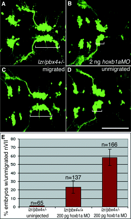
Fig. 4 Loss of one copy of lazarus/pbx4 enhances effects of hoxb1a morpholinos on nVII motor neuron migration. (A–D) Confocal images of live embryos at 36 hpf. Brackets indicate the positions of facial (nVII) motor neurons in each panel. (A) lzr/pbx4+/- facial motor neurons migrate normally as in lzr/pbx4+/+ embryos (see Fig. 1E). (B) A total of 2 ng of hoxb1aMO blocks presumptive facial motor neuron migration in 95% of injected, lzr/pbx4+/+ embryos. (C, D) Classes of phenotypes observed after injecting 10-fold less (200 pg) hoxb1a morpholino: (C) “migrated” and (D) “unmigrated.” (E) A summary of the average percentage of lzr/pbx4+/+ and lzr/pbx4+/- embryos with unmigrated r4-derived motor neurons following injection of 200 pg hoxb1aMO. Error bars represent standard deviations across five independent experiments. The value “n” indicates the total number of embryos represented in these experiments. Chi-square tests on data from individual experiments demonstrate statistical significance to the differences observed between lzr/pbx4+/+ and lzr/pbx4+/- embryos (P < 0.01). Scale bar, 100 μm.
Reprinted from Developmental Biology, 253(2), Cooper, K.L., Leisenring, W.M., and Moens, C.B., Autonomous and nonautonomous functions for Hox/Pbx in branchiomotor neuron development, 200-213, Copyright (2003) with permission from Elsevier. Full text @ Dev. Biol.

