Image
Figure Caption
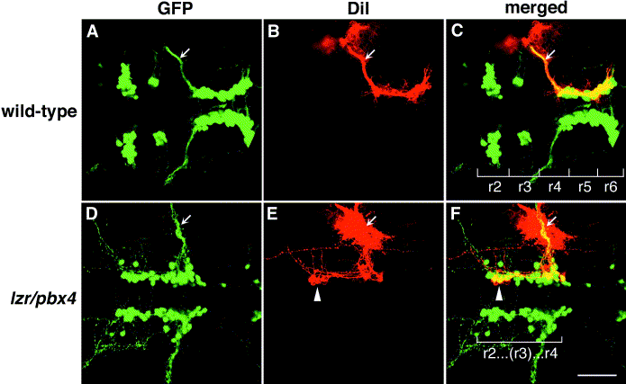
Fig. 2 Motor axons from r2 and r3 of lzr/pbx4-/- embryos misroute through the r4 exit point with the facial (nVII) nerve. Anterior is to the left in all panels. DiI was applied to the nVII nerve root distal to its r4 exit point (arrow) in fixed, 36-hpf embryos to retrogradely label nVII cell bodies within the hindbrain. (A–C) DiI fills cells located primarily in r5 and r6 of wild-type embryos. (D–F) DiI applied to the nVII root of lzr/pbx4-/- embryos fills cells in r4 as well as cells located in r2 and r3 (arrowhead in E and F). Scale bar, 50 μm.
Acknowledgments
This image is the copyrighted work of the attributed author or publisher, and
ZFIN has permission only to display this image to its users.
Additional permissions should be obtained from the applicable author or publisher of the image.
Reprinted from Developmental Biology, 253(2), Cooper, K.L., Leisenring, W.M., and Moens, C.B., Autonomous and nonautonomous functions for Hox/Pbx in branchiomotor neuron development, 200-213, Copyright (2003) with permission from Elsevier. Full text @ Dev. Biol.

