
|
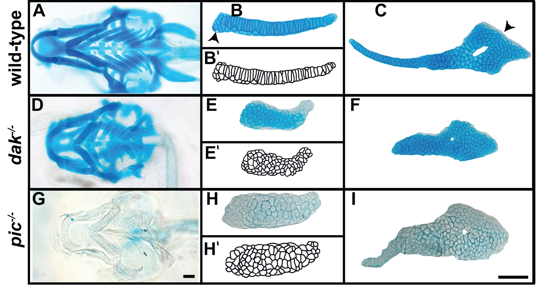
Fig. 1 dak-/- and pic-/- larvae have similar cartilage morphogenesis phenotypes.
Skeletal preparations of wild-type (A–C), dak-/- (D–F) and pic-/- (G–I) at day 6 reveal the shape of the cartilaginous skeleton as well as chondrocyte morphology. Ventral views of the head show that the cartilage elements of dak-/- and pic-/- fish are shorter and thicker than wild-type (A,D,G). Dissected cartilage laid flat show a complete lack of chondrocyte flattening and intercalation in skeletal elements from dak-/- and pic-/- larvae (B,E,H ceratobranchial 1; C,F,I hyosymplectic). Arrowheads in B and C indicate regions that lack stacking in wild-type embryos. Alcian Blue staining at pH1.0 (HCl 0.1N) does not stain pic-/- cartilage (G,H,I). Camera lucida drawings of chondrocytes in wild-type and mutant larvae (B′,E′,H′). Scale bars = 50μM.