
|
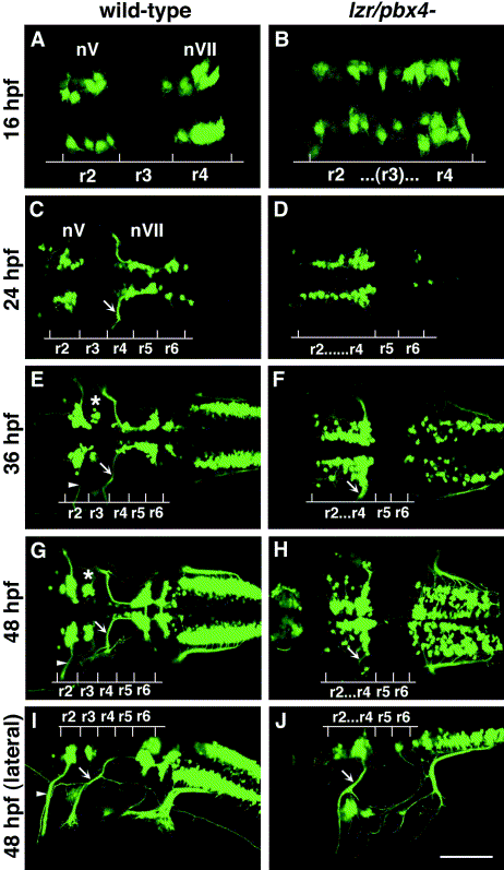
Fig. 1 Confocal images of Isl1-GFP expression in live wild-type (left column) and lzr/pbx4-/- (right column) embryos. Anterior is to the left in all panels. All images are a dorsal view, except (I) and (J) which are lateral views. (A, B) The onset of GFP expression in trigeminal (nV) motor neurons in r2 and facial (nVII) motor neurons in r4 occurs at 16 hpf. In lzr/pbx4-/- embryos, motor neurons differentiate prematurely in r3. (C, D) By 24hpf in wild-type embryos, nVII cell bodies have migrated into r5 and r6, and axons leave r4 (arrow). In lzr/pbx4-/- embryos, presumptive nVII cells have not migrated posteriorly. (E, F) nV motor neurons in r3 appear by 36 hpf in wild-type embryos (asterisk). Arrows mark the nVII motor nerve exiting in r4; arrowhead in (E) marks the nV motor nerve exiting in r2. (G, H) By 48 hpf in wild-type embryos, nVII motor neurons have completed their migration into r6 and r7, while in lzr/pbx4-/- embryos, presumptive nVII motor neurons remain in r4. Labeling is as in (E) and (F). (I, J) Lateral views of 48-hpf embryos show a strong reduction of the nV nerve (arrowhead in I) in lzr/pbx4-/- embryos accompanied by a thickening of the nVII nerve (arrow in I, J). Scale bar, 50 μm in (A) and (B); 100 μm in (C–J).
Reprinted from Developmental Biology, 253(2), Cooper, K.L., Leisenring, W.M., and Moens, C.B., Autonomous and nonautonomous functions for Hox/Pbx in branchiomotor neuron development, 200-213, Copyright (2003) with permission from Elsevier. Full text @ Dev. Biol.