
|
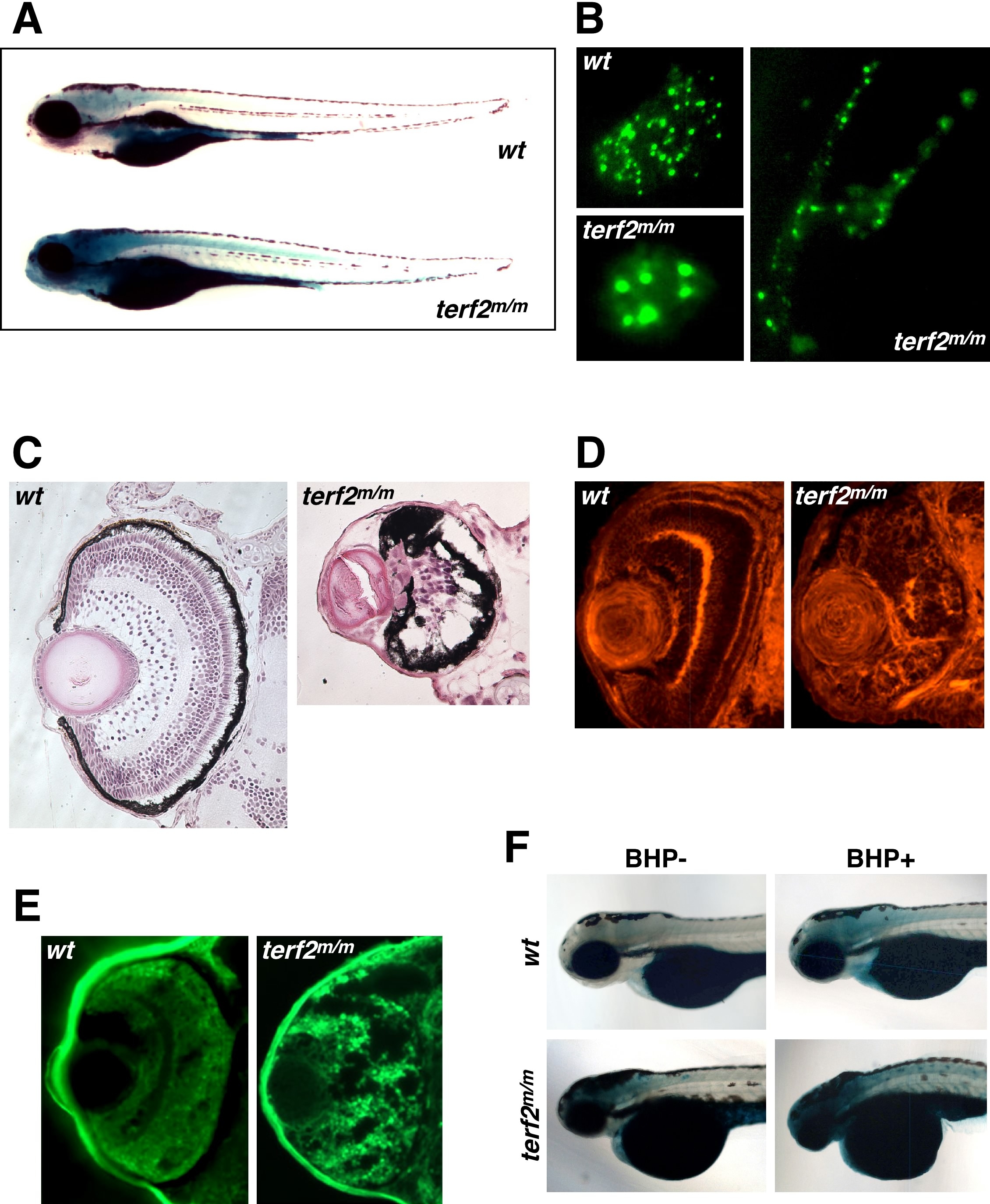
Fig. 4
Mutant terf2 animals with high SA-β-gal activity and retinal neurodegenerative phenotypes.
(A) 4.5-day old (4.5 dpf) homozygous terf2m/m zebrafish larvae show high SA-β-gal activity, particularly in the brain and spinal cord, having the small eyes and head (lower image), compared with wild-type (upper image). (B) Abnormally enlarged telomere speckles (lower left panel) and aberrant nuclear shapes (right panel) can be observed in homozygous terf2m/m embryos, compared with normal telomere speckles in a nuclear of the wild-type embryo (upper left panel) at 5 dpf. (C) H&E staining of transverse sections through the retinas of homozygous terf2m/m (right panel) and wild-type zebrafish embryos (left panel) at 5 dpf. (D) Embryonic zebrafish retinas were stained with phalloidin to visualize the actin filaments in the plexiform layers, which revealed obvious structural defects in the homozygous terf2m/m mutant (right panel) compared with a wild-type sibling (left panel) at 3 dpf. (E) Neurodegeneration in the retina was histologically detected by performing Fluoro-Jade B staining in homozygous terf2m/m mutant (right panel), but was not evident in the wild-type embryo (left panel) at 2 dpf. (F) Homozygous terf2 mutant embryos and wild-type controls were exposed to 350 μM BHP from 6 hpf to 4 dpf. Enhanced SA-β-gal staining with a more severe morphology in eyes and heads are observed in BHP-treated terf2 mutant embryos at 4 dpf (lower right panel).