
Fig. 1
- ID
- ZDB-IMAGE-081021-129
- Publication
- Thisse et al., 2003 - Spatial and temporal expression patterns of selenoprotein genes during embryogenesis in zebrafish
- All Figures
- Figures for Thisse et al., 2003
|
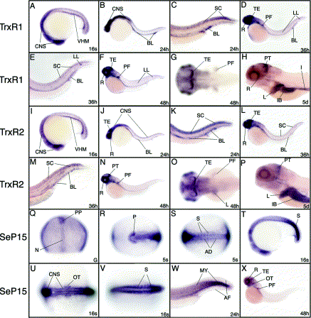
Fig. 1 Expression patterns of the thioredoxin reductase (A–H), glutathione/thioredoxin reductase (I–P) and 15 kDa selenoprotein (Q–R) during development in zebrafish embryos. Development stage is indicated in the right lower corner (G, gastrula; 5s, 5 somites; 16s, 16 somites or in hours post-fertilization). AD, adaxial cells; AF, axial fin fold; BL, blood; CNS, central nervous system; I, intestine; IB, intestinal bulb; L, liver; LL, lateral line; MY, myotomes; N, notochord; OT, otic vesicle; P, polster; PF, pectoral fins; PP, prechordal plate; PT, posterior tectum; R, retina; S, somites; SC, spinal chord; TE, tectum; VHM, ventral hematopoetic mesoderm.
Reprinted from Gene expression patterns : GEP, 3(4), Thisse, C., Degrave, A., Kryukov, G.V., Gladyshev, V.N., Obrecht-Pflumio, S., Krol, A., Thisse, B., and Lescure, A., Spatial and temporal expression patterns of selenoprotein genes during embryogenesis in zebrafish, 525-532, Copyright (2003) with permission from Elsevier. Full text @ Gene Expr. Patterns