
|
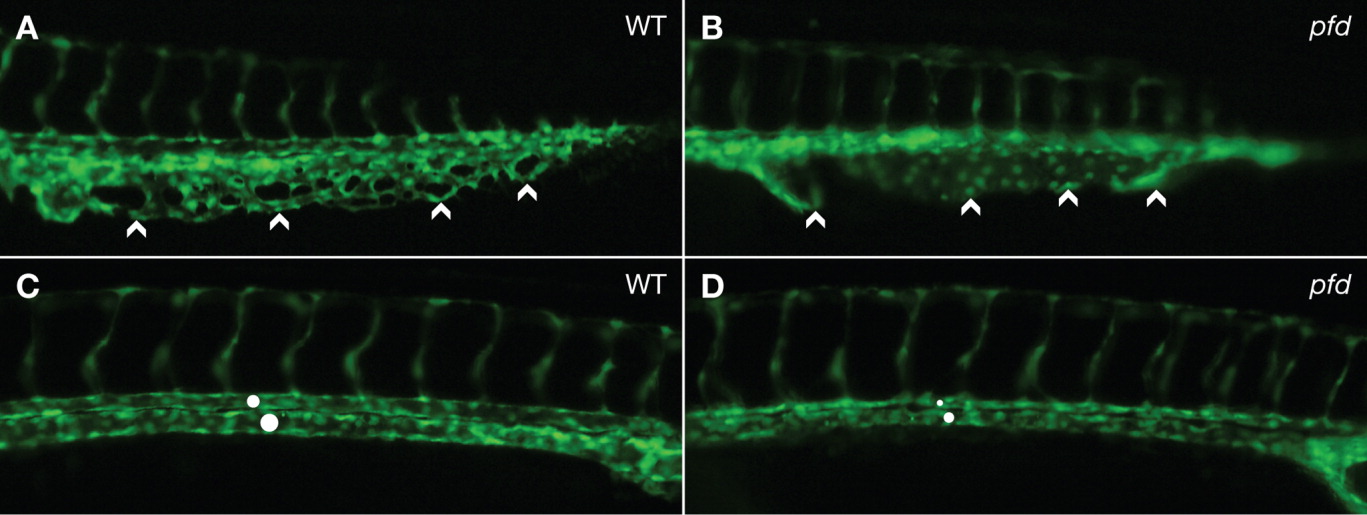
Fig. 2 The pfdgw1 mutation disrupts venous plexus and axial vessel formation. A-D: pfdgw1 was crossed into a fli1:EGFP transgenic line to allow visualization of endothelial cells. A: The caudal vein of wild-type embryos has a well-formed venous plexus (arrowheads). B: The caudal vein of pfdgw1 mutants has lost its characteristic reticular pattern, and endothelial cells are disorganized (arrowheads). C,D: Dorsal aorta (upper circle) and cardinal vein (lower circle) in a wild-type embryo (C) and a pfdgw1 mutant (D) demonstrating reduced axial vessel diameters in the mutant (D, circles). Embryos were photographed at 30 hours postfertilization (hpf; C,D) and 35 hpf (A,B).