
|
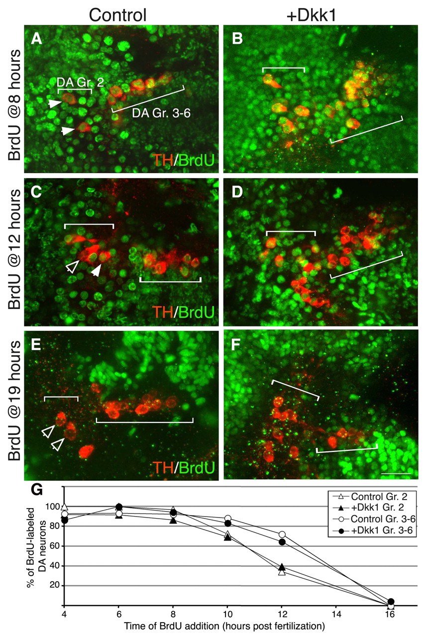
Fig. 5 Dkk1 does not delay cell cycle exit. (A-F) Projected confocal z-stack images of wild-type (WT; A,C,E) and dkk1- injected (B,D,F) zebrafish embryos at 48 hpf. Proliferating DA progenitors were labeled by BrdU at the indicated time points followed by immunofluorescence staining with antibodies against tyrosine hydroxylase (TH) and BrdU. The positions of DA group 2 (Gr. 2) and groups 3-6 (Gr. 3-6) are indicated. Solid arrowheads indicate TH+ BrdU+ double-positive neurons; open arrowheads indicate TH+ neurons with no nuclear BrdU staining. (G) Plot of the percentage of proliferating group 2 (Gr. 2; triangles) and group 3-6 (Gr. 3-6; circles) DA progenitor cells as a function of time in WT (white symbols) and dkk1-injected (black symbols) populations. Scale bar: 25 μm.