
|
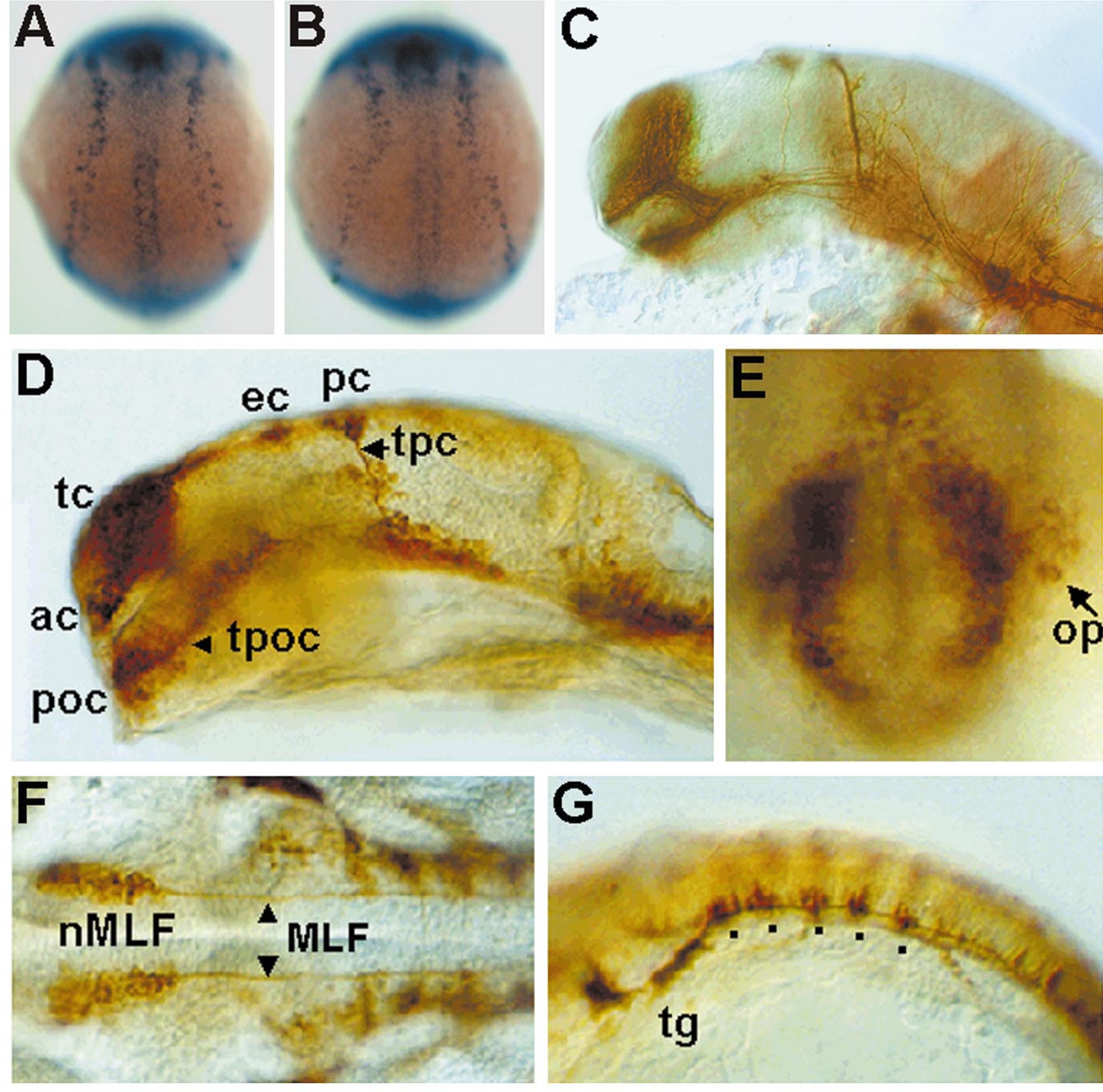
Fig. 8 Temporal and spatial expression pattern of HuC-GFP minigene in the homozygotic transgenic zebrafish embryos. Expression patterns of GFP (A) and HuC (B) mRNA transcripts detected by whole mount in situ hybridization using synthetic antisense RNAs. Dorsal view of 11 hpf embryos for GFP (A) and HuC (B). (C) Lateral view, expression of acetylated α-tubulin detected by whole mount immunostaining (Sigma mouse Monoclonal, clone 6-11B-1). Expression of GFP labeled by anti-GFP polyclonal antibody in 24 hpf embryos (D–G). (D) Lateral view; expressions of GFP were detected in the telencephalic cluster (tc), anterior commissure (ac), epiphysial cluster (ec), posterior commissure (pc), tract of posterior commissure (tpc), postoptic commissure (poc), and tract of the postoptic commissure (tpoc). (E) Anterior view, expression of GFP in the olfactory placodes. (F) Dorsal view, expression of GFP in the nucleus of the medial longitudinal fasciculus (nMLF) and medial longitudinal fasciculus (MLF). (G) Lateral view of hindbrain; expression of GFP in the trigeminal ganglion (tg) and rhombomeres (v) in the hindbrain.
Reprinted from Developmental Biology, 227(2), Park, H.-C., Kim, C.-H., Bae, Y.-K., Yee, S.-Y., Kim, S.-H., Hong, S.-K., Shin, J., Yoo, K.-W., Hibi, M., Hirano, T., Miki, N., Chitnis, A.B., and Huh, T.-L., Analysis of upstream elements in the HuC promoter leads to the establishment of transgenic zebrafish with fluorescent neurons, 279-293, Copyright (2000) with permission from Elsevier. Full text @ Dev. Biol.