
|
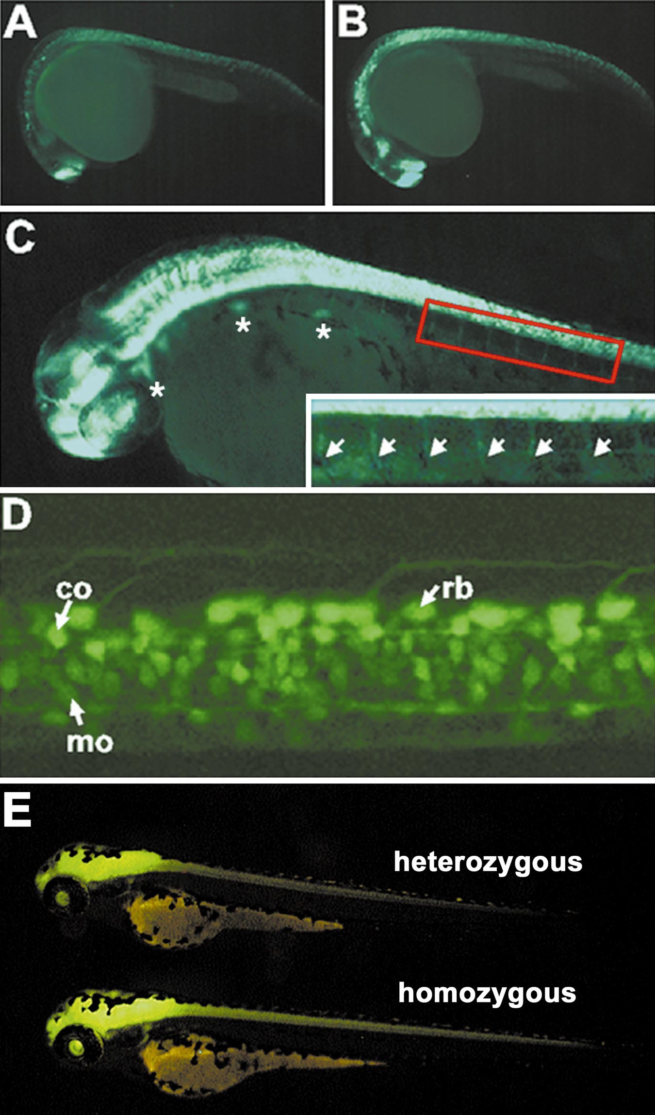
Fig. 7 GFP fluorescence of neuron in living transgenic zebrafish. The neuronal cells of transgenic zebrafish lines permanently expressing the HuC-GFP minigene (10.7 kb) were analyzed by the fluorescence microscopy. Lateral views of GFP fluorescence detected in the neurons of 24 hpf heterozygotic (A) and homozygotic (B) transgenic embryos. (C) Cranial ganglia are highlighted by asterisks. Ventral motor roots in the boxed area are indicated by arrows. (D) Lateral view of spinal cord of 60 hpf transgenic zebrafish embryo. The Rohon-Beard cells (rb), commissural neurons (co), and primary motorneurons (mo) are visualized by the GFP fluorescence in living transgenic zebrafish embryo. (A, B, C) GFP fluorescence detected by stereomicroscope; (D) Pseudo-color image of GFP fluorescence analyzed by laser confocal microscopy.
Reprinted from Developmental Biology, 227(2), Park, H.-C., Kim, C.-H., Bae, Y.-K., Yee, S.-Y., Kim, S.-H., Hong, S.-K., Shin, J., Yoo, K.-W., Hibi, M., Hirano, T., Miki, N., Chitnis, A.B., and Huh, T.-L., Analysis of upstream elements in the HuC promoter leads to the establishment of transgenic zebrafish with fluorescent neurons, 279-293, Copyright (2000) with permission from Elsevier. Full text @ Dev. Biol.