
Image
|
Figure Caption
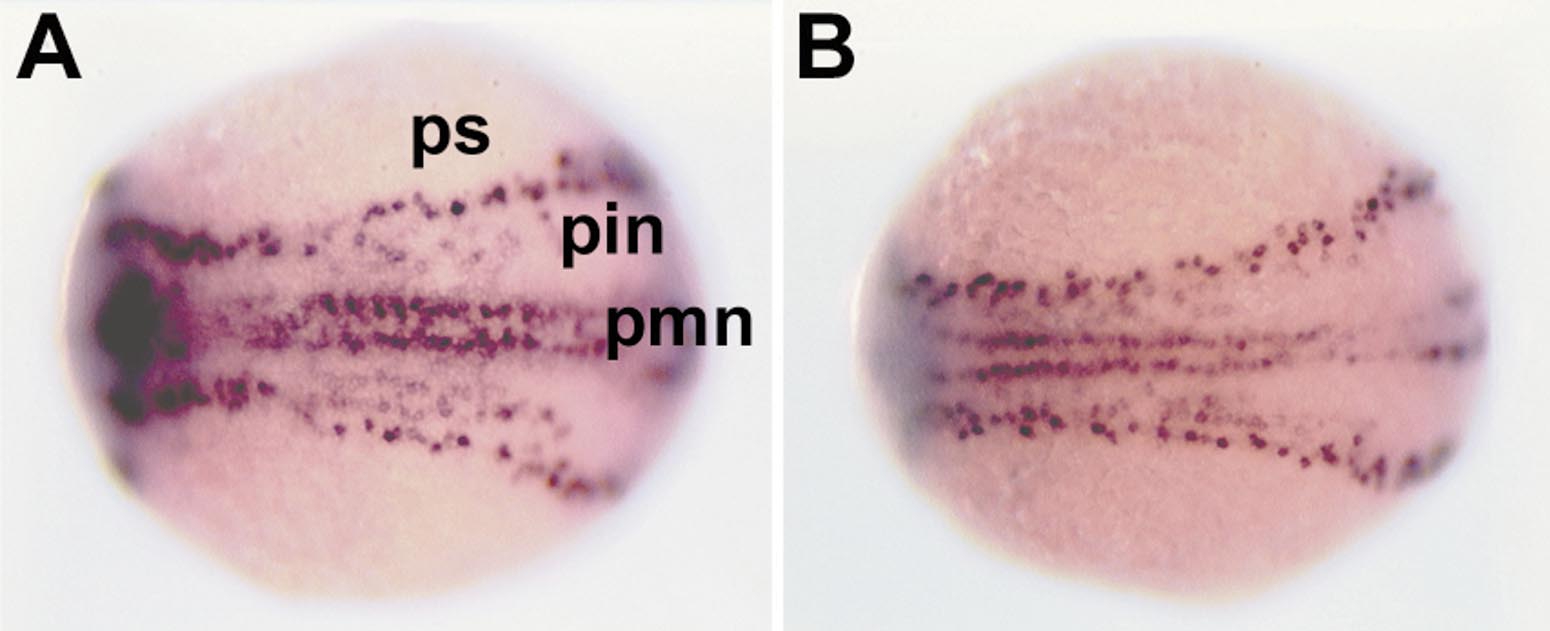
Fig. 1 Comparison of HuC and DeltaB mRNA expression in the neural plate at the 3-somite stage. HuC (A) is expressed in isolated cells in a pattern very similar to that of DeltaB (B), which is expressed in neuronal precursor cells. Dorsal views, anterior to the left. ps, primary sensory neuron; pin, primary intermediate neuron; pmn, primary motor neuron.
Acknowledgments
This image is the copyrighted work of the attributed author or publisher, and
ZFIN has permission only to display this image to its users.
Additional permissions should be obtained from the applicable author or publisher of the image.
Reprinted from Developmental Biology, 227(2), Park, H.-C., Kim, C.-H., Bae, Y.-K., Yee, S.-Y., Kim, S.-H., Hong, S.-K., Shin, J., Yoo, K.-W., Hibi, M., Hirano, T., Miki, N., Chitnis, A.B., and Huh, T.-L., Analysis of upstream elements in the HuC promoter leads to the establishment of transgenic zebrafish with fluorescent neurons, 279-293, Copyright (2000) with permission from Elsevier. Full text @ Dev. Biol.