
|
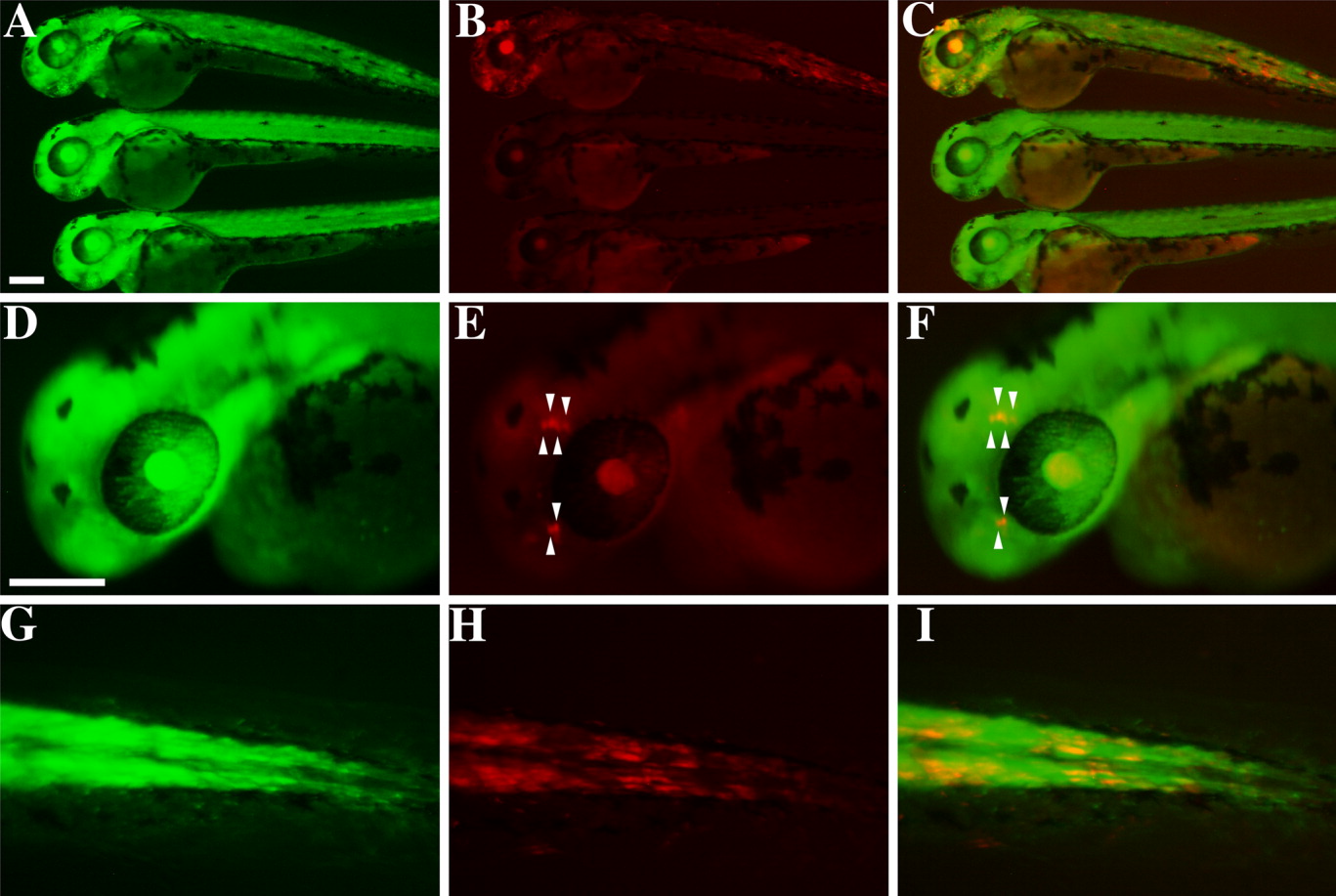
Fig. 3 Cre-mediated recombination at the single cell level is detected by color conversion from green to red in the 2 days postfertilization (dpf) G2R embryos injected with 8xHSE-Cre. A,D,G: Left column, GFP. B,E,H: Center, RFP. C,F,I: Right, merged images. A,B,C: The 8xHSE-Cre injected and heat-shocked embryo (top) showed mosaic expression of RFP induced by transient transgenic Cre expression, while injected and unheated (center) and uninjected and unheated (bottom) embryos displayed the background level of red signals only. D,E,F: High magnification of the color conversion in the head of an 8xHSE-injected and heat-shocked embryo shows four-cell and two-cell clusters of RFP-positive cells (indicated by arrowheads). G,F,I: High magnification of the color conversion in the trunk-tail region of an 8xHSE-injected, heat-shocked embryo indicates RFP-positive cells in clusters of various sizes. Scale bars = 200 μm.