
|
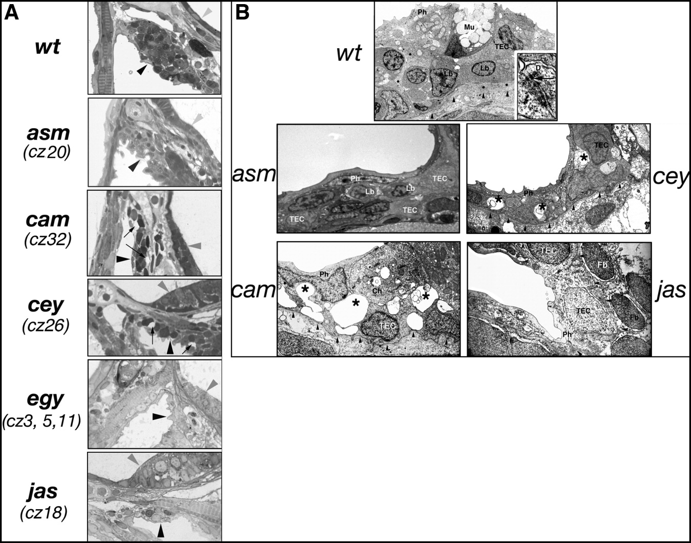
Fig. 5 Abnormal thymic architecture in mutant larvae. A: Light microscopic examination of wild-type 7 dpf thymus shows a heterogeneous, cellular thymus (dark arrowhead). Epithelium of the otic capsule is indicated by grey arrowhead. Thymus of cey and cam mutant larvae shows reduction in thymic epithelial cell layers and empty spaces (arrows). Magnification was x100 for all panels. B:Electron microscopy of 7 dpf wt thymus (top panel) shows a heterogeneous population of cells. Arrowheads indicate basement membrane. Insert (x54,000) in right lower corner shows desmosomes (D, arrow) between thymic epithelial cells. Occasional lymphoblasts were only found in asm mutants. Empty spaces suggesting degeneration in the thymic epithelial cells were encountered in cey and cam mutants (asterisks). Magnification was x4,000 for all panels. Ch, chloride cell, a salt-producing cell occurring in the epithelium of pharyngeal cavity of many fish species; Fb, fibroblast; Lb, lymphoblast; Mu, mucine producing cell; Ph, pharyngeal epithelial cell; TEC, thymic epithelial cell.