
Image
|
Figure Caption
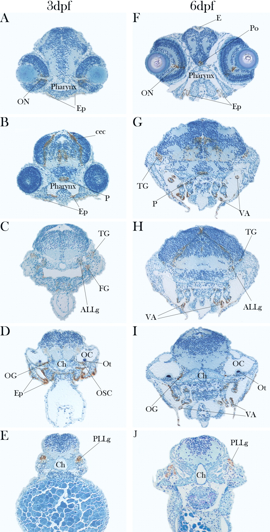
Fig. 5 Immunostaining of the CLGY786/spry1 line, cross-sections of anterior regions show domains where YFP protein is expressed. A-E: At 3 hours postfertilization (hpf). F-J: At 6 hpf. ALLg, anterior lateral line ganglion; cec, cerebellar commissure; Ch, chorda dorsalis; E, epiphysis; Ep, epithelium; FG, facial ganglion; mlf, medial longitudinal fascicle; OC, otic capsule; OG, otic ganglion; OSC, otic support cells; Ot, otolith; Po, preoptic region; TG, trigeminal ganglion; VA, visceral arches. For other abbreviations, see Figure 2 and 3.
Figure Data
Acknowledgments
This image is the copyrighted work of the attributed author or publisher, and
ZFIN has permission only to display this image to its users.
Additional permissions should be obtained from the applicable author or publisher of the image.
Full text @ Dev. Dyn.