
Image
|
Figure Caption
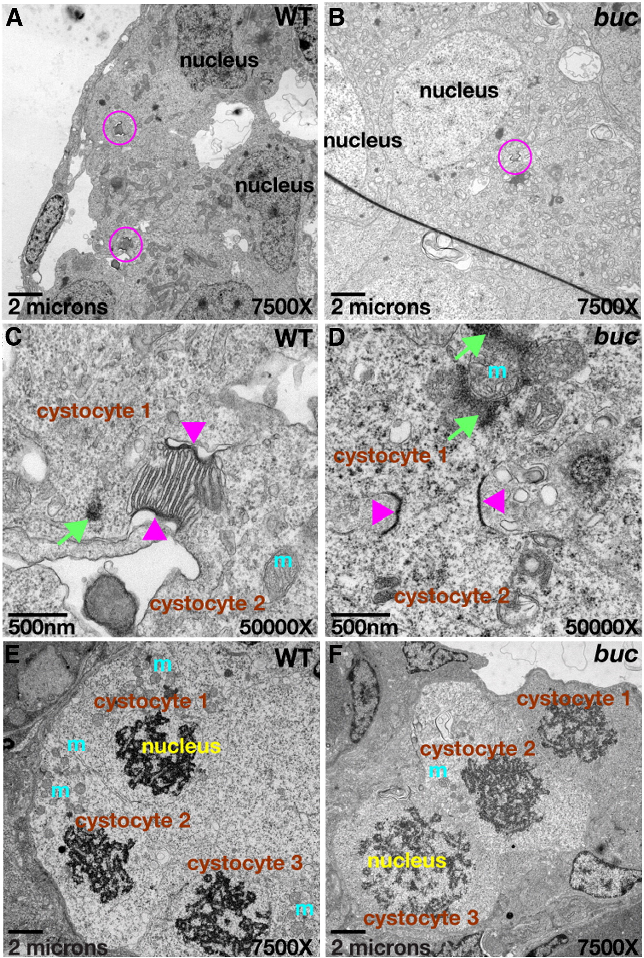
Fig. 5 Cytoplasmic bridges connect cystocytes in zebrafish. Ring canal-like cytoplasmic bridges (pink circles) connect primary oocytes in a cyst (cystocytes) in panel A wild-type and (B) buc mutants. (C, D) 50,000x TEM images of cysts from (A) and (B), mitochondrial cement (green arrows) and mitochondria (m) are near the cytoplasmic bridges (pink arrowheads) in wild-type and mutants. (E, F) Synchronous cyst division in wild-type and buc mutant cysts; 7500x TEM images.
Figure Data
Acknowledgments
This image is the copyrighted work of the attributed author or publisher, and
ZFIN has permission only to display this image to its users.
Additional permissions should be obtained from the applicable author or publisher of the image.
Reprinted from Developmental Biology, 321(1), Marlow, F.L., and Mullins, M.C., Bucky ball functions in Balbiani body assembly and animal-vegetal polarity in the oocyte and follicle cell layer in zebrafish, 40-50, Copyright (2008) with permission from Elsevier. Full text @ Dev. Biol.