
Image
|
Figure Caption
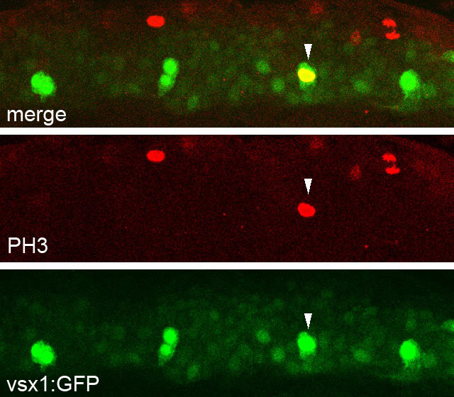
Fig. S2 Mitotic cells are included among the vsx1-GFP cell population. Anti-PH3 (phospho-Histone H3) immunohistochemical staining in a Tgvsx1:GFP embryo at 17 hpf. A vsx1-GFP cell (arrowhead) is also positive for PH3.
Figure Data
Acknowledgments
This image is the copyrighted work of the attributed author or publisher, and
ZFIN has permission only to display this image to its users.
Additional permissions should be obtained from the applicable author or publisher of the image.
Full text @ Development