
|
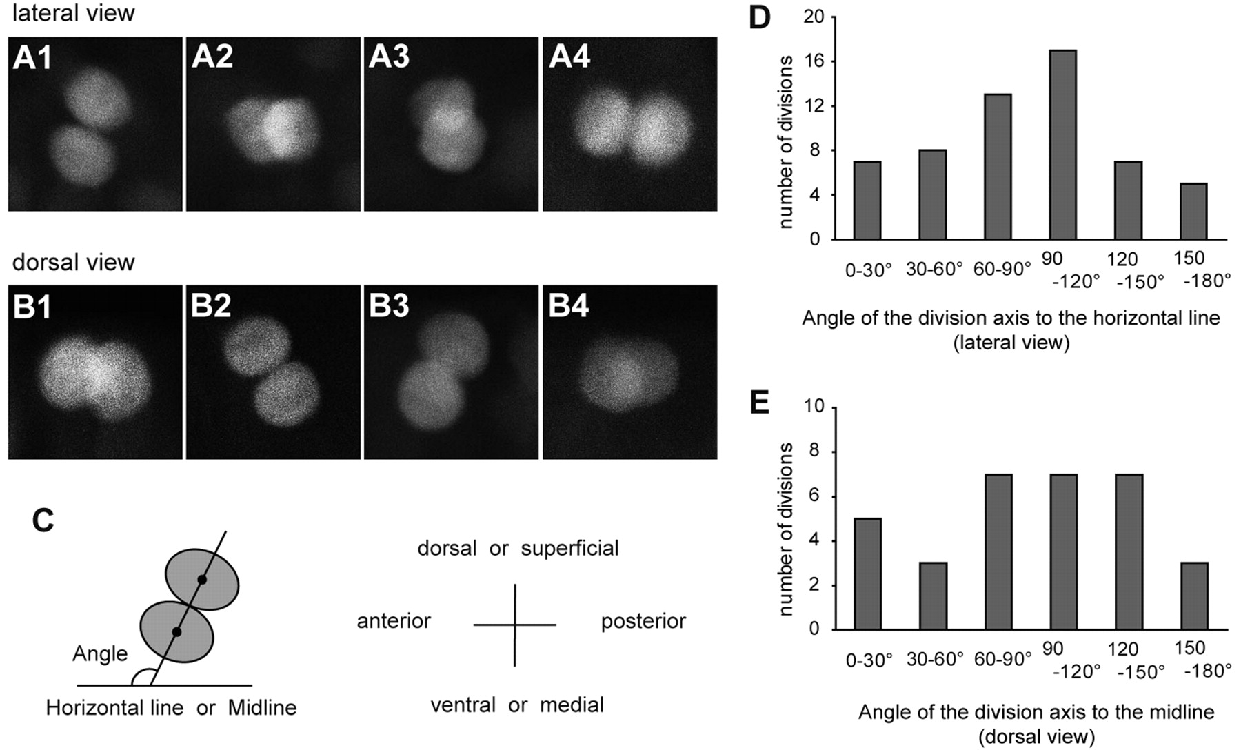
Fig. 3 Division axes of p2 intermediate progenitors are not fixed. (A,B) Confocal stacked images of vsx1-GFP sibling cells immediately after division. Embryos were analyzed at around 16-17 hpf. (A1-A4) Lateral views. Dorsal is towards the top. (B1-B4) Dorsal views. Superficial is towards the top. (C) Schematic of the method for division axis measurements. For each division, a line connecting the centers of the two sister cell (projection of division axis) was drawn. The angle of this line to the horizontal line (lateral views) or to the midline (dorsal views) was measured. (D,E) Quantification of the division angle.