
|
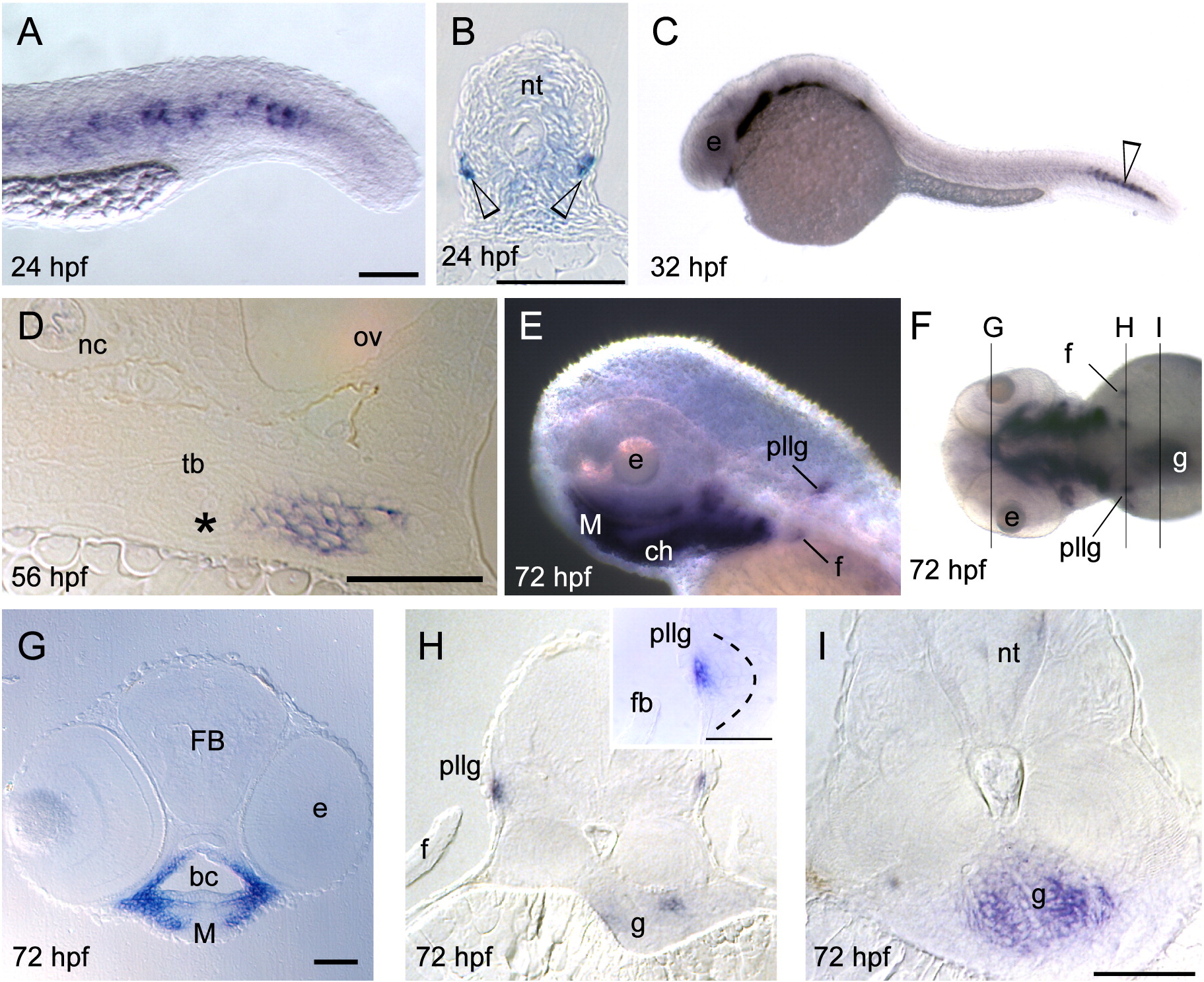
Fig. S2 Whole-mount in situ hybridization of barx1 expression. (A–B) 24 hpf, (C) 32 hpf, (D) 56 hpf, and (E–I) 72 hpf. (A) Tail expression. (B) Cross-section of the trunk, open arrowheads indicates barx1 expression. (D) Coronal section of the fifth branchial arch adjacent to the tooth bud primordium (asterisk). (E) Lateral view of pharyngeal arch expression, posterior lateral line ganglia (pllg), and fin (f) expression domains. (F) Dorsal view indicating coronal serial sections in panels (G, H and I). (G) Expression in the arch mesenchyme of the jaw. (H) Cross-section through the pllg. (H inset) Higher magnification of expression in the posterior lateral line ganglia; dotted line outlines ganglion. (I) Cross-section through the gut wall (g). f, fin; FB, forebrain; M, Meckel's cartilage; nc, notochord; nt, neural tube; ov, otic vesicle; tb, tooth bud. Scale bar: (A) 100 μm, (B, D, G, H inset and I) 50 μm.
Reprinted from Developmental Biology, 321(1), Sperber, S.M., and Dawid, I.B., barx1 is necessary for ectomesenchyme proliferation and osteochondroprogenitor condensation in the zebrafish pharyngeal arches, 101-110, Copyright (2008) with permission from Elsevier. Full text @ Dev. Biol.