
|
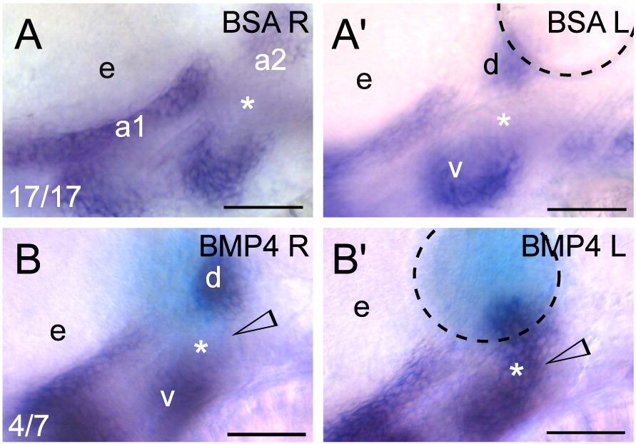
Fig. 9 Exogenous BMP signaling influences barx1 expression within the zebrafish pharyngeal arches. (A, B) Whole-mount in situ hybridization of barx1 in the left (bead implanted) and right (control) side of single embryos. Right-side images were flipped for easier comparison. (A, A′) Control BSA white bead, (B, B′) BMP4 blue bead. Anterior is to the left; d and v, dorsal and ventral expression domains in the second arch; e, eye. Arrowhead indicates sites of barx1 misexpression; asterisk indicates intermediate region, normally devoid of barx1 expression at this stage. Number of affected embryos over total embryos with successful bead implantation is indicated in the panels A, B. Black dashed line outlines the bead. Scale bar: 50 μm.
Reprinted from Developmental Biology, 321(1), Sperber, S.M., and Dawid, I.B., barx1 is necessary for ectomesenchyme proliferation and osteochondroprogenitor condensation in the zebrafish pharyngeal arches, 101-110, Copyright (2008) with permission from Elsevier. Full text @ Dev. Biol.