
|
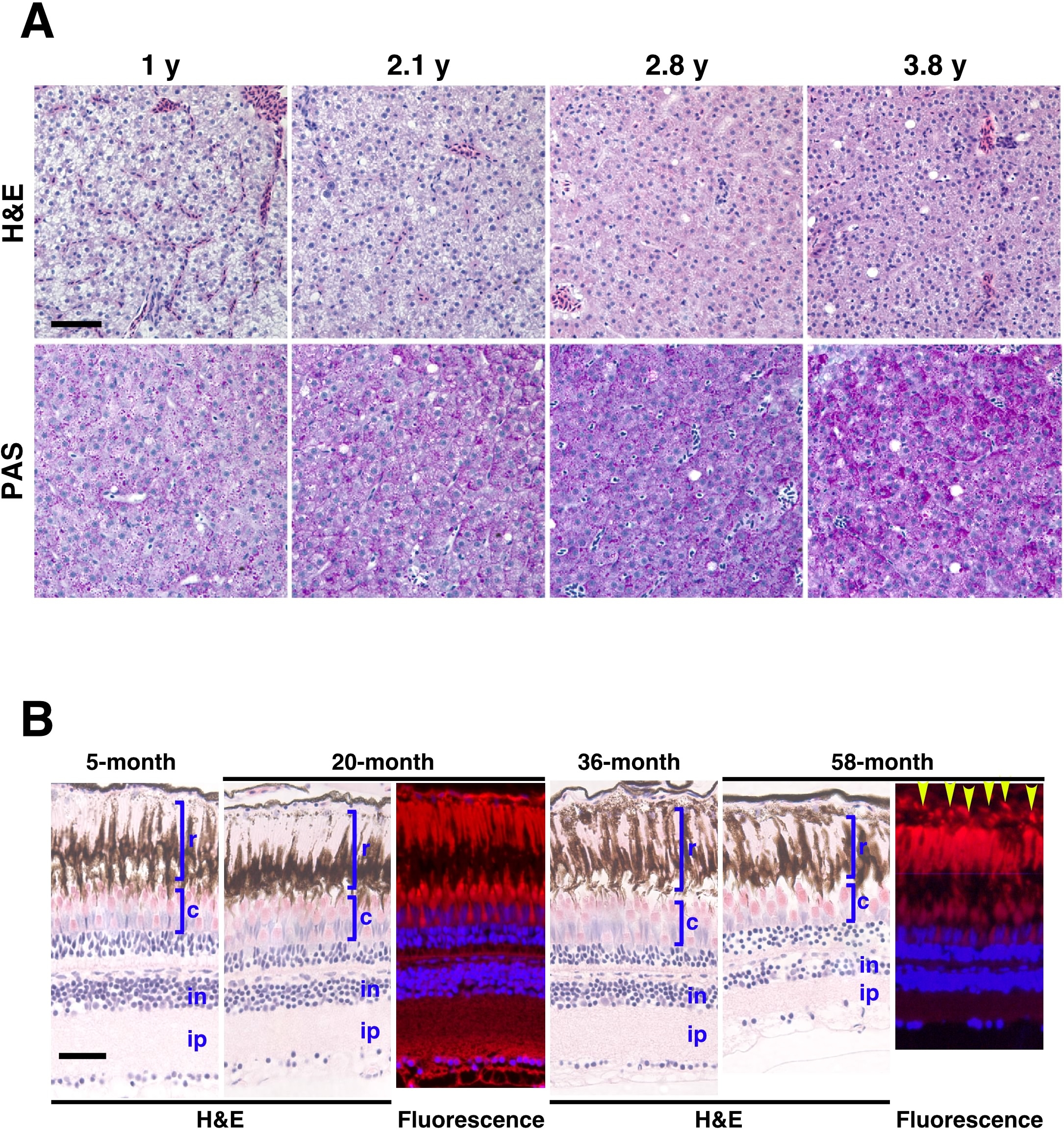
Fig. S7 Liver and eye histology in the adult zebrafish with age. (A) Liver sections of 1 y, 2.1 y, 2.8 y, and 3.8 y wild-type zebrafish were stained by H&E or PAS staining. Hepatocyte density and eosinophilic staining can be seen to increase with age by H&E staining. PAS-positive staining for lipofuscin also shows increased levels of this biomarker in aging liver tissue. (B) Eye sections of 5-month, 20-month, 36-month, and 58-month old wild-type fish were stained by H&E. In the light adapted retina, the rods (r) sit distally to the cones (c); ‘in’ indicates the inner nuclear layer, and ‘ip’ indicates the inner plexiform layer. Processes from the pigment epithelium (PE) extend between the outer segments of the rods. Aged (58-month old) wild-type fish show increased drusen-like accruals (yellow arrows) with autofluorescence in the RPE (lower right panel), compared with younger (20-month old) wild-type fish (upper right panel). (Scale bar: 100 μm.)