
|
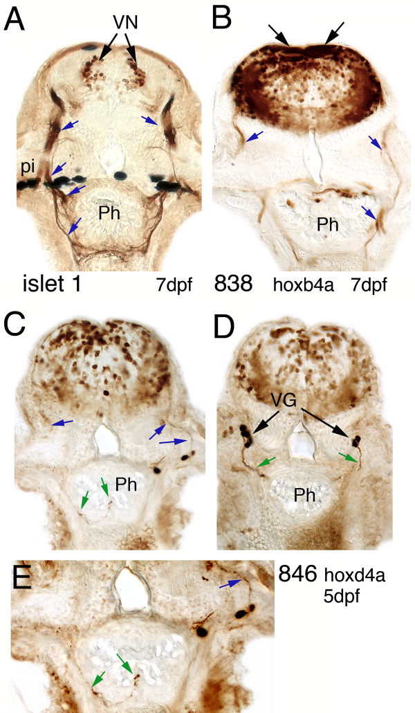
Fig. 4 Sections through anti-GFP (islet1 GFP transgenic line) and anti-YFP (hoxb4a and hoxd4a enhancer detection lines) immunostained larvae. A: Islet 1 expression at 7 dpf in the vagal nucleus (dorsal black arrows) and the vagal visceromotor nerve (blue arrows). B: Hoxb4a regulated YFP expression in a broad hindbrain domain and in a subpopulation of the vagal nerve (blue arrows). The black arrows point to neuropithelial cells, which are YFP positive. C-E: Hoxd4a-YFP expression in 5-dpf larvae. C: A section anterior of the first somite. The blue arrows point to weakly stained fibers of the vagal nerve exiting the hindbrain. Vagal ganglion cells and a fine fiber innervating the pharynx (green arrows) are also YFP positive. D: One section further posterior to C with YFP-labeled vagal ganglion cells that project towards the pharynx (green arrows). E: A magnification of C with focus on the fine fibers that innervate the pharynx. Arrows follow the same color code as in C and D. Ph, pharynx; pi, pigment.