
Image
|
Figure Caption
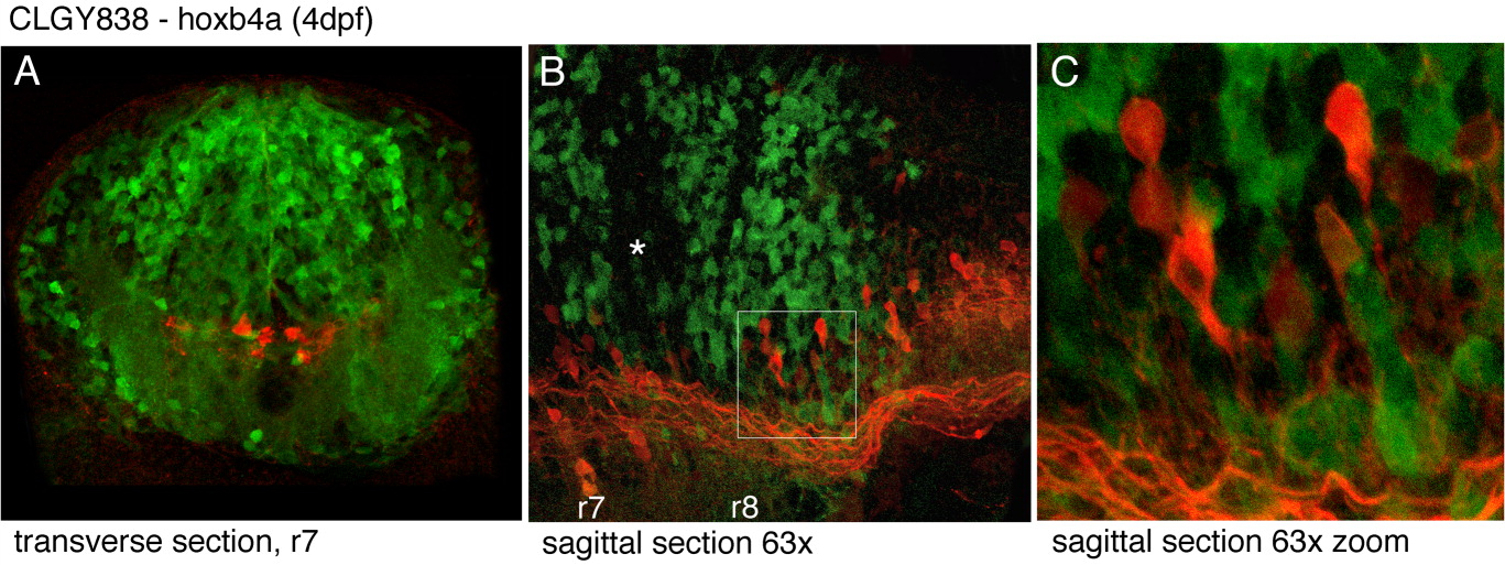
Fig. 7 The reticulospinal formation was labeled in CLGY838 larvae at 4 dpf and cryosections through these specimen were double immunostained for Biotin and YFP. YFP-positive cells are shown in green, the rhodamin-dextan labeled reticulospinal neurons are red. Hoxb4a-YFP cells did not overlap with reticulospinal neurons. A: Transverse section at first somite level. B: Sagittal section through the medial hindbrain. The asterisk marks a region where YFP-positive neurons are absent. C: Magnification of inset from sagittal section.
Figure Data
Acknowledgments
This image is the copyrighted work of the attributed author or publisher, and
ZFIN has permission only to display this image to its users.
Additional permissions should be obtained from the applicable author or publisher of the image.
Full text @ Dev. Dyn.