
|
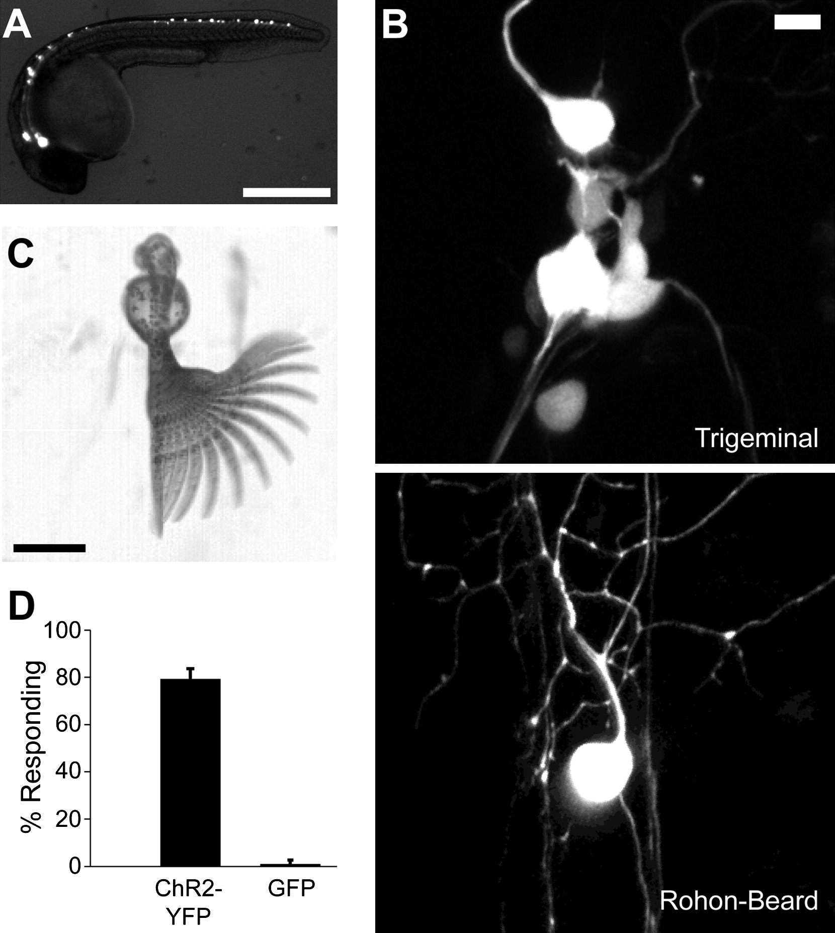
Fig. 1 Photoactivation of ChR2 in Zebrafish Somatosensory Neurons Triggers Escape Behaviors
(A) Lateral view of a 24 hpf embryo expressing ChR2-YFP and EGFP in touch-sensitive Rohon-Beard and trigeminal neurons. Anterior is at left, and dorsal is at top. The scale bar represents 100 μm.
(B) Maximum-intensity Z projections of two-photon stacks showing ChR2-YFP and EGFP expression in trigeminal (top panel) and Rohon-Beard (bottom panel) neurons at 24 hpf. The scale bar represents 10 μm.
(C) Time-series projection of a ChR2-YFP-expressing embryo performing an escape in response to illumination at 488 nm. Images were acquired at 500 fps. The scale bar represents 100 μm.
(D) Percentage of experimental (Isl1::Gal4-VP16::UAS-E1b::ChR2-YFP, UAS::GFP) and control (Isl1::Gal4-VP16, UAS::GFP) embryos showing light-evoked escape behaviors. Data are mean ± SEM across three (ChR2) and two (control) clutches of 50–100 injected embryos. Experimental: 79% ± 4%, n = 149 embryos; control: 1% ± 1%, n = 66 embryos.