
|
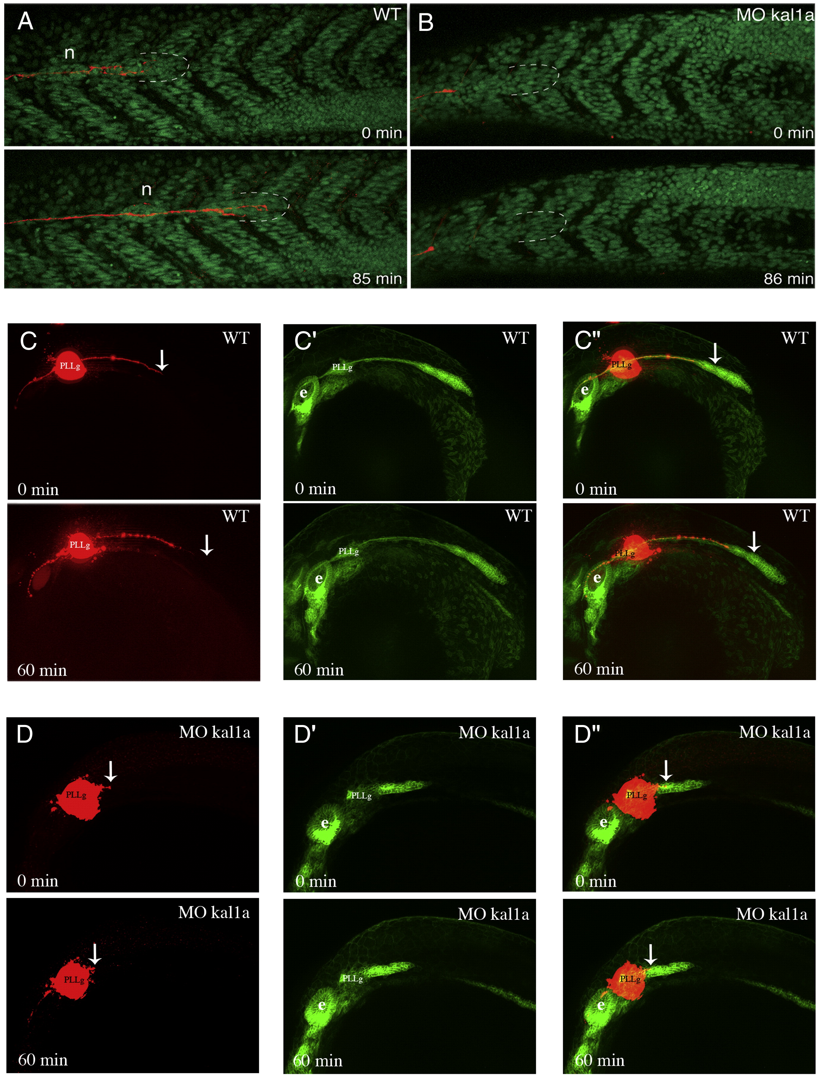
Fig. 5 Kal1a inactivation severely compromises PLLP migration as shown by DiI labelling of the axon of PLL sensory neurons in H2A.F/Z::GFP (A and B) and ClaudinB::GFP (C to D″) embryos. In vivo imaging of PLLP migration by time-lapse confocal microscopy from 32 to 33.5 hpf in a H2A.F/Z::GFP embryo (A) and a H2A.F/Z::GFP MO kal1a (0.5 mM) morphant (B), and apotome microscopy from 26 to 27 hpf in a ClaudinB::GFP embryo (C to C″) and a ClaudinB::GFP MO kal1a (0.5 mM) morphant (D to D″) following DiI injection into the PLLP ganglion. Dashed lines: PLLP leading edge; n: recently deposited neuromast. PLLg: posterior lateral line ganglion, e: ear, WT: wild-type. Arrows indicate the tip of the PLLP nerve. Anterio-posterior axis is from left to right.
Reprinted from Developmental Biology, 320(2), Yanicostas, C., Ernest, S., Dayraud, C., Petit, C., and Soussi-Yanicostas, N., Essential requirement for zebrafish anosmin-1a in the migration of the posterior lateral line primordium, 469-479, Copyright (2008) with permission from Elsevier. Full text @ Dev. Biol.