
|
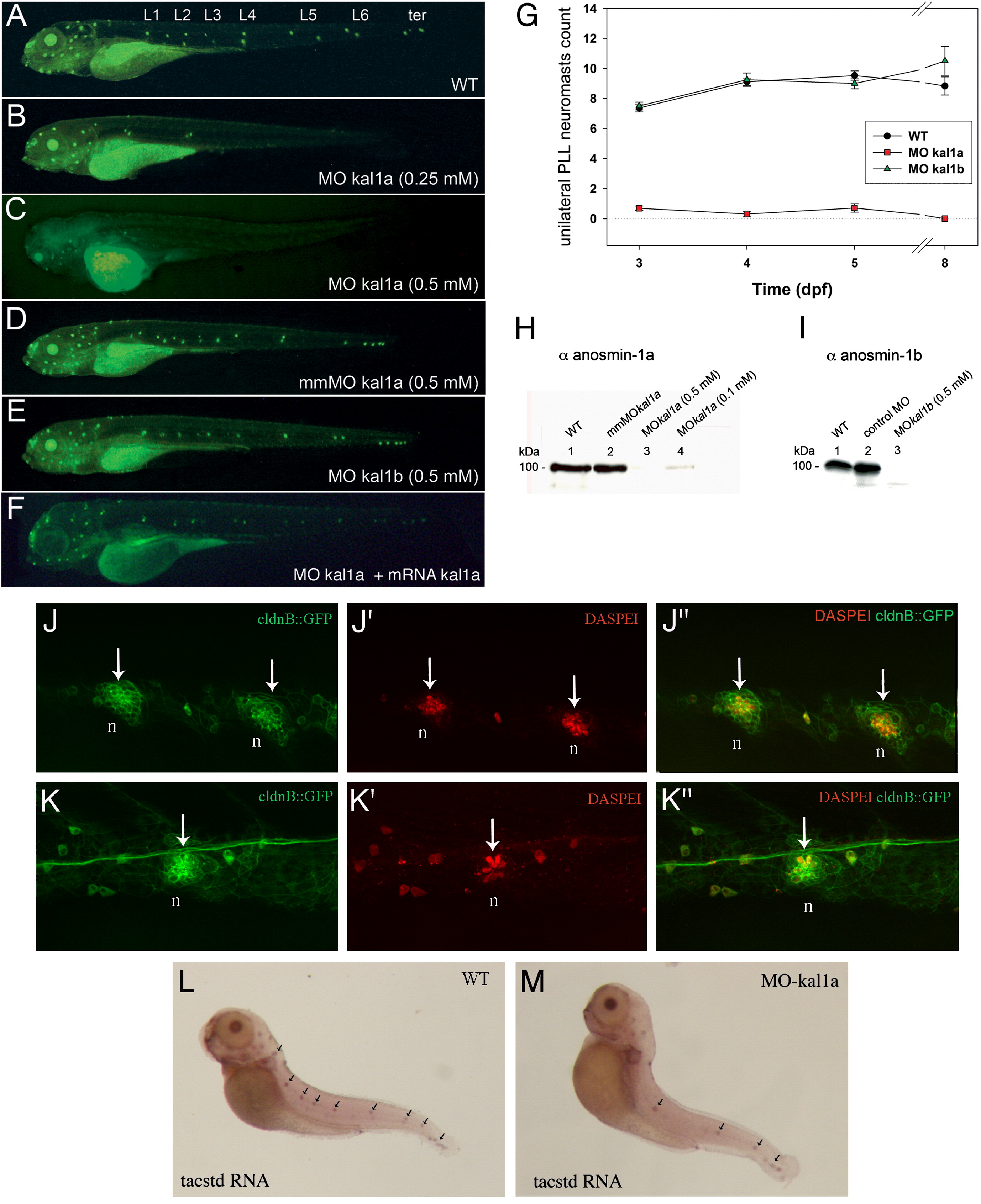
Fig. 2 Morpholino-mediated kal1a, but not kal1b, inactivation impairs PLL neuromast deposition as visualized by DASPEI staining. Wild-type (WT) embryo (A), MO kal1a (0.25 mM) (B) or (0.5 mM) (C), mmMO kal1a (0.5 mM) (D), MO kal1b (0.5 mM) morphants (E) and MO kal1a (0.5 mM) morphant co-injected with MO kal1a-insensitive kal1a RNA (1 μM) (F). Time course of neuromast deposition in WT embryos (n = 48), kal1a (0.5 mM) (n = 53) and kal1b (0.5 mM) morphants (n = 45) from 3 to 8 dpf (G). Values are means ± s.e.m. Western blot analysis of anosmin-1a accumulation (H) in 3 dpf WT embryos (1), mmMO kal1a (0.5 mM) (2), MO kal1a (0.5 mM) (3) and (0.1 mM) morphants (4) and anosmin-1b accumulation (I) in 3 dpf WT embryos (1), MO control (2) and MO kal1b (0.5 mM) morphants (3). Neuromast organization in wild-type embryos (J–J″ and L) and intermediate MO kal1a (0.25 mM) morphant (K–K″ and M) visualized by DASPEI staining of hair cells on ClaudinB::GFP embryos (J–K″) and in situ detection of tacstd transcripts in neuromast support cells (L, M). L1–L6: neuromasts lined up along the flanks of embryos; ter: neuromasts clustered in the tail region. Arrows indicate neuromasts. n: neuromast.
Reprinted from Developmental Biology, 320(2), Yanicostas, C., Ernest, S., Dayraud, C., Petit, C., and Soussi-Yanicostas, N., Essential requirement for zebrafish anosmin-1a in the migration of the posterior lateral line primordium, 469-479, Copyright (2008) with permission from Elsevier. Full text @ Dev. Biol.