
Image
|
Figure Caption
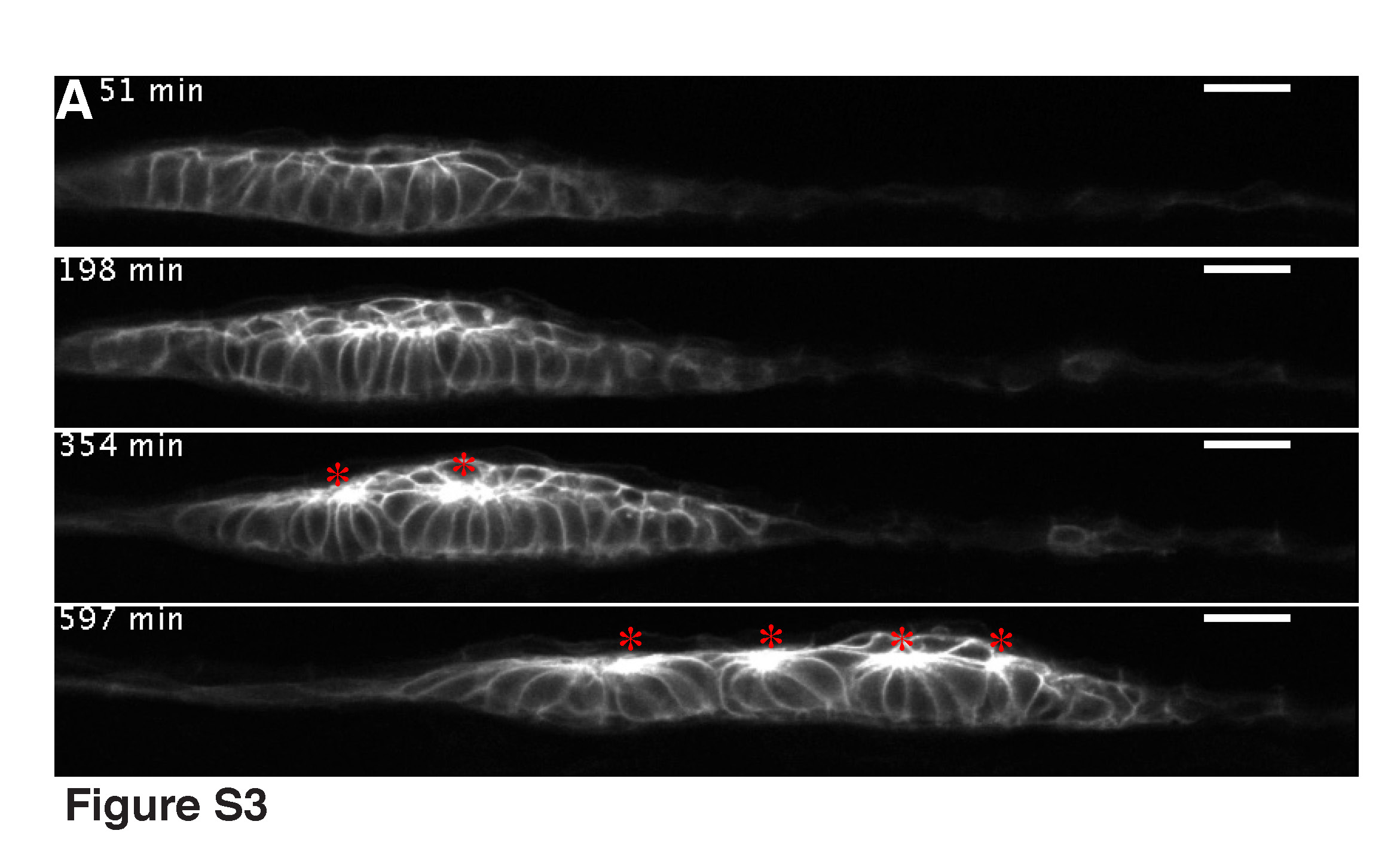
Fig. S3 Time-lapse analysis showing epithelialized cells snapping together to form rosettes in the second phase of washout. The embryo was pre-treated with SU5402, washed for 5 hours and mounted side-on. Asterisks indicate the formed rosettes (see also Movie 7).
Acknowledgments
This image is the copyrighted work of the attributed author or publisher, and
ZFIN has permission only to display this image to its users.
Additional permissions should be obtained from the applicable author or publisher of the image.
Full text @ Development