
|
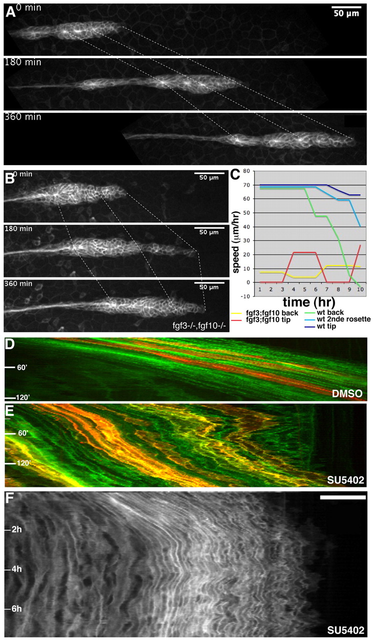
Fig. 4 The primordium migrates in an uncoordinated manner in the absence of FGF activity. (A) Images from a time-lapse movie of wild-type migration, velocity 69 μm/hour. (B) Images from a time-lapse movie of a cldnb:gfp fgf3-/-;fgf10-/- embryo taken over 6 hours, showing the uncoordinated migration of the front and the back, average velocity 11 μm/hour. (C) Graph showing the speed of migration of tip (dark blue), the second rosette (blue) and the back (green) of the wild-type primordium, and of the tip (red) and the back (yellow) of the double mutant primordium, over a 10-hour period. (D) Kymograph of wild-type migration; all cells move at a similar speed, as indicated by the parallel lines of the kymograph. (E) Kymograph of a 3-hour movie of an SU5402-treated mosaic wt primordium showing the uncoordinated migration of cells within the primordium. (F) Kymograph of an 8-hour movie of a SU5402-treated wt primordium showing the back and forth movement of the primordium before it stops.