
|
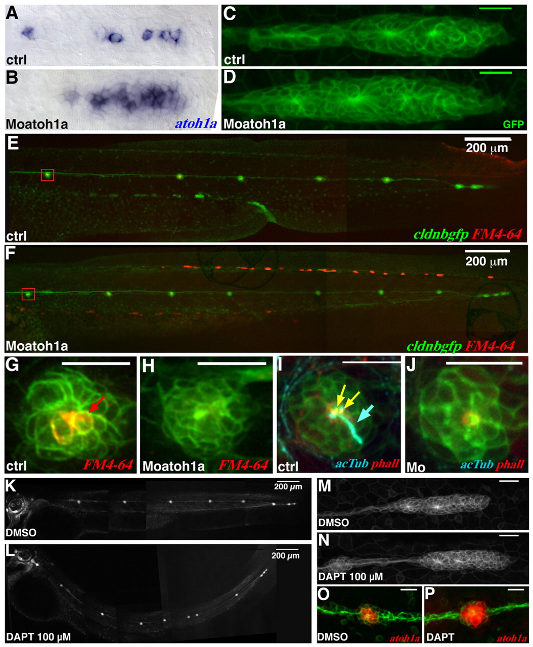
Fig. 3 Hair cell specification is not required for rosette assembly. (A,B) atoh1a expression is expanded in MoAtoh1a-injected embryos (B) compared with control embryos (A). (C,D) Live cldnb:gfp control (C) and MoAtoh1a-injected (D) embryos at 36 hpf. (E,F) The same embryos as in C,D imaged at 2.5 dpf, the number and position of neuromasts is within normal range. (G,H) Close-up view of a neuromast (red box in E,F), with hair cells labeled with FM4-64. Differentiated hair cells are present in wild type (red arrow) but absent in MoAtoh1a-injected embryos. (I,J) Loss of hair cells was confirmed by using an anti-acetylated tubulin antibody that labels the kinocilium (blue, blue arrow). (K,L) The primordium in embryos treated with 100 μm DAPT shows a normal pattern of neuromast deposition. (M,N) Primordia in DAPT and control embryos are indistinguishable. (O,P) The loss of restricted atoh1a expression in deposited neuromasts confirms the efficacy of DAPT treatment. Scale bars: 20 μm (unless otherwise stated).