
|
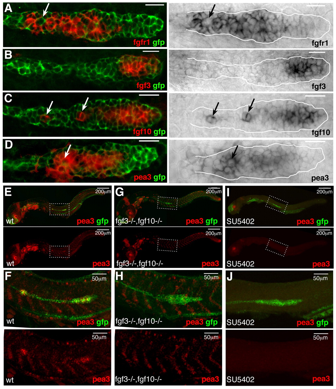
Fig. 1 FGF signaling is active in the migrating primordium. (A-D) Confocal images of the primordium in 36-hpf cldnb:gfp embryos labelled with a GFP antibody and indicated in situ hybridization probes. Right panels are transmission images of NBT-BCIP stainings, which were inverted and overlaid with GFP (left). Arrows point to fgf10-expressing cells (C), or to cells with reduced expression of fgfr1 (A) or pea3 (D). (E-J) Fluorescent images of 36-hpf cldn:bgfp wild-type (E,F), fgf3;fgf10 double mutant (G,H) and SU5402-treated (I,J) embryos labelled with a pea3 probe (red) and GFP (green). F,H and J are close-ups of the primordium in E,G and I (white dashed frames). Anterior is to the left. Scale bars: 20 μm (unless stated otherwise).