
|
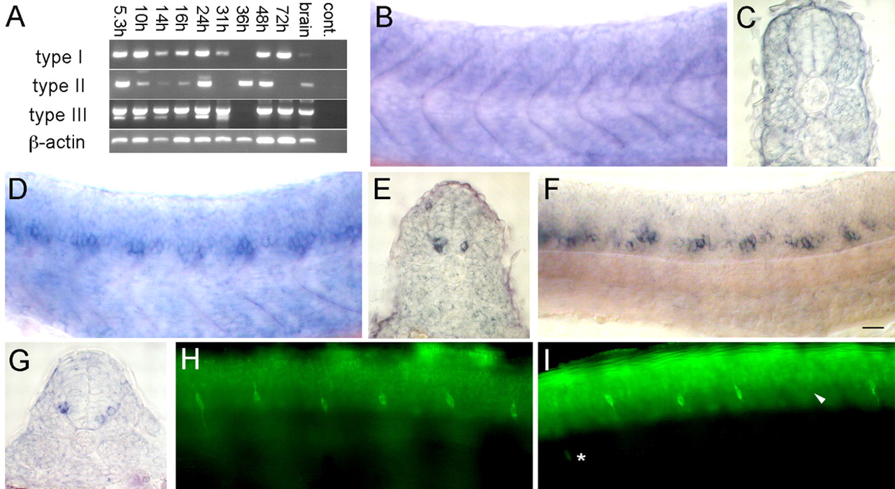
Fig. 5 nrg1 alone is not required for DRG formation. (A) RT-PCR showing expression of each nrg1 isoform at various developmental stages and in the adult brain. (B,D,F) Expression patterns of each isoform at 24 hpf shown in whole-mount. (C,E,G) Cross section of expression pattern of each isoform respectively. (B,C) Type I is expressed weakly in somites. (D,E) Type II and (F,G) type III are expressed strongly in ventral spinal cord neurons. (E,F) Elavl labeling at 2 dpf. (H,I) Control MO-injected embryos (H) had segmental DRG neurons. nrg1 MO-injected embryos (I) had only a slight decreased in DRG neuron number; arrowhead indicates absent DRG, asterisk shows mislocalized DRG. Scale bar: 40 μm.