
|
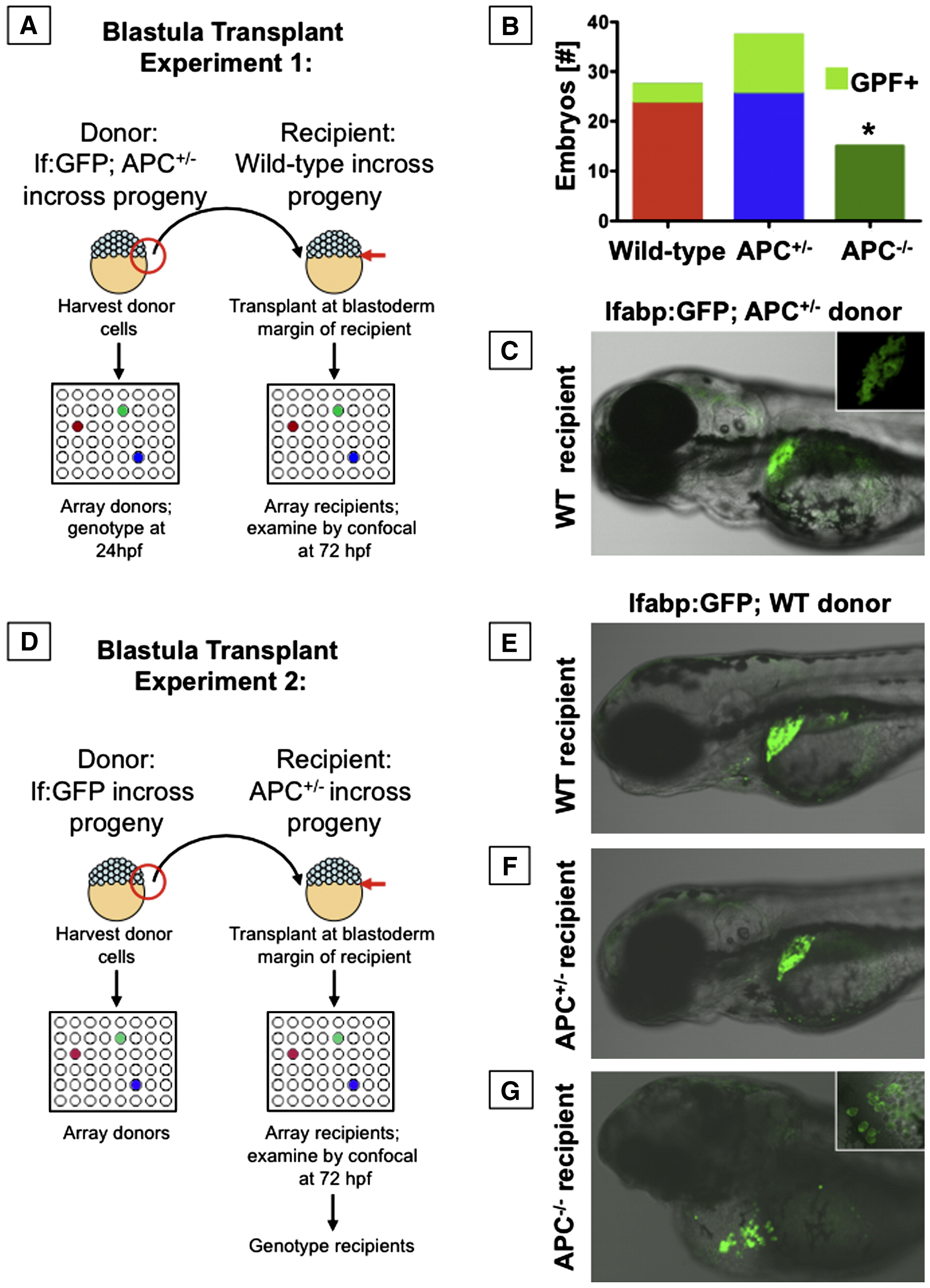
Fig. 6 APC has cell autonomous effects on endoderm development. Blastula transplant experiments; embryos were analyzed at 72 hpf. (A) Schematic depiction of blastula transplant experiments with each APC genotype as donor. (B) Graphic summary of the number of recipient embryos that received each donor genotype; the fraction of embryos that showed donor contribution to the liver is highlighted in light green. No APC-/- donor cells contributed to liver formation (Fisher's exact, p = 0.025). (C) Mosaic livers showed green hepatocytes interspersed with unlabeled cells. (D) Schematic depiction of blastula transplant experiments with different APC genotypes as recipients. (E, F) lfabp:GFP donor cells transplanted into both wild-type or APC+/- hosts gave rise to mosaic livers. (G) In an APC-/- host, lfabp:GFP hepatocytes developed, but could not rescue liver development. The inset shows formation of chains of hepatocytes near the heart and around the yolk sac. Significant differences are indicated with an asterisk (∗).
Reprinted from Developmental Biology, 320(1), Goessling, W., North, T.E., Lord, A.M., Ceol, C., Lee, S., Weidinger, G., Bourque, C., Strijbosch, R., Haramis, A.P., Puder, M., Clevers, H., Moon, R.T., and Zon, L.I., APC mutant zebrafish uncover a changing temporal requirement for wnt signaling in liver development, 161-174, Copyright (2008) with permission from Elsevier. Full text @ Dev. Biol.