
|
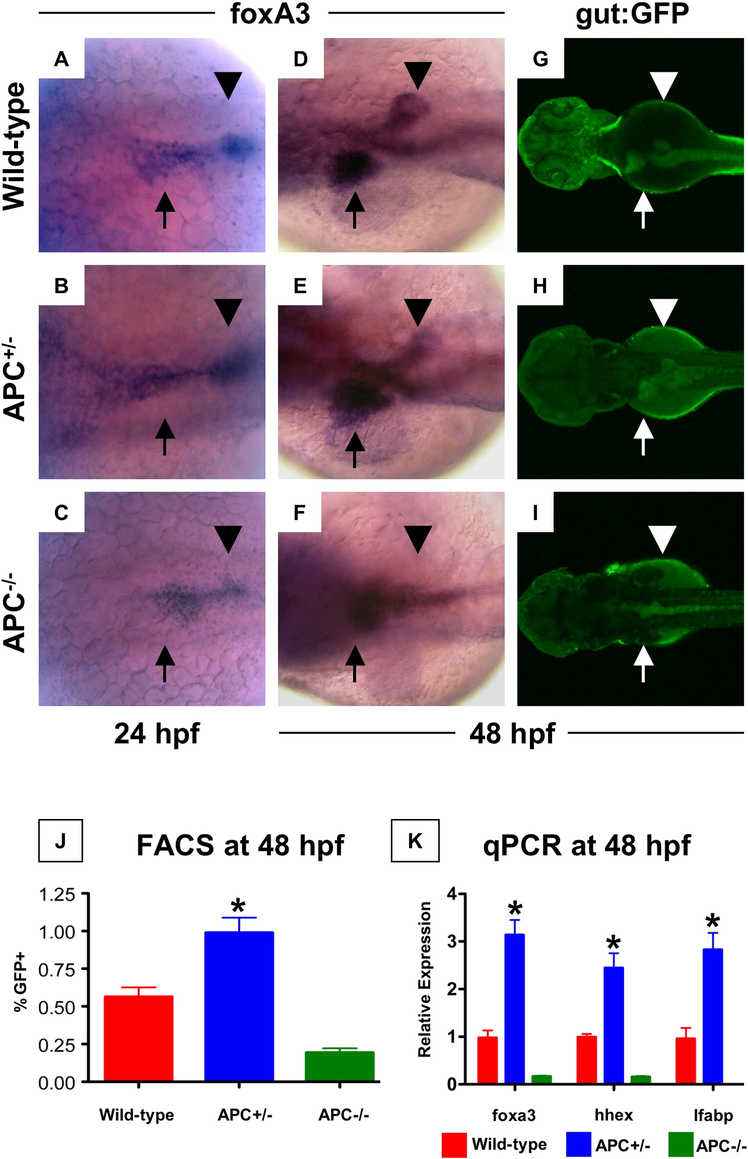
Fig. 4 APC loss affects endodermal and hepatic progenitor cells. (A–C) In situ hybridization for foxa3 revealed changes in endodermal progenitor organization in APC mutant embryos as early as 24 hpf. (D–F) By 48 hpf, this led to a progressive increase in hepatic and corresponding decrease in pancreatic buds in the APC+/- embryos (35 altered/49 scored), while the APC-/- embryos failed to develop an organized endodermal pattern (23/25). (G–I) In vivo confocal fluorescence imaging of gut:GFP transgenic embryos at 48 hpf revealed similar effects on endodermal patterning. (J) FACS analysis demonstrated a doubling in the number of gut:GFP+ (green gate) endodermal progenitor cells in APC+/- embryos compared to wild-type controls; GFP+ cells were severely diminished in APC-/- embryos (APC+/+ 1.56 ± 0.063%; APC+/- 0.99 ± 0.099%; APC-/- 0.19 ± 0.089% of 20,000 cells analyzed; ANOVA, n = 10/genotype, p < 0.00001). (K) qPCR analysis confirmed the increased expression levels of foxa3, hhex and lfabp in APC+/- (blue) embryos and the depressed/absent expression in APC-/- (green) embryos at 48 hpf (ANOVA, n = 10/category, p < 0.05).
Reprinted from Developmental Biology, 320(1), Goessling, W., North, T.E., Lord, A.M., Ceol, C., Lee, S., Weidinger, G., Bourque, C., Strijbosch, R., Haramis, A.P., Puder, M., Clevers, H., Moon, R.T., and Zon, L.I., APC mutant zebrafish uncover a changing temporal requirement for wnt signaling in liver development, 161-174, Copyright (2008) with permission from Elsevier. Full text @ Dev. Biol.