
Image
|
Figure Caption
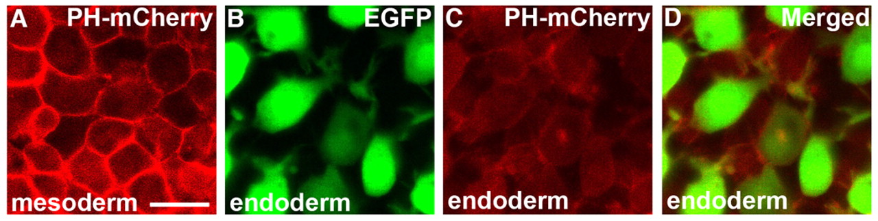
Fig. 7 Specific subcellular localization of PH-mCherry is not observed in migrating endodermal cells. (A-D) Confocal analysis of PH-mCherry localization in mesodermal and endodermal cells at the 90% epiboly stage. In mesodermal cells, PH-mCherry is significantly localized at the cell membrane (A), but this is not observed in endodermal cells (B-D). Scale bar: 20 μm.
Acknowledgments
This image is the copyrighted work of the attributed author or publisher, and
ZFIN has permission only to display this image to its users.
Additional permissions should be obtained from the applicable author or publisher of the image.
Full text @ Development