
Image
|
Figure Caption
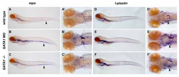
Fig. S1 The gata1 MO-injected (B, B′) and vlt mutant embryos (C, C′) have an increased number of cells expressing mpo at 4 dpf compared to wild-types (A, A′). Expression of mpo in the tail (arrowheads) is especially affected. The number of cells expressing l-plastin at 4 dpf also increased in the gata1 MO-injected (E, E′) and vlt mutant embryos (F, F′) compared to wild-types (D, D′) particularly in the head regions (B′–F′).
Figure Data
Acknowledgments
This image is the copyrighted work of the attributed author or publisher, and
ZFIN has permission only to display this image to its users.
Additional permissions should be obtained from the applicable author or publisher of the image.
Reprinted from Developmental Cell, 8(1), Galloway, J.L., Wingert, R.A., Thisse, C., Thisse, B., and Zon, L.I., Loss of gata1 but not gata2 converts erythropoiesis to myelopoiesis in zebrafish embryos, 109-116, Copyright (2005) with permission from Elsevier. Full text @ Dev. Cell