
Image
|
Figure Caption
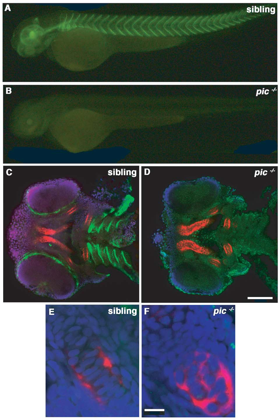
Fig. S2 The 10E4 epitope is not expressed in the developing cartilage. Wildtype (A,C,E) and pic-/- (B,D,F) localisation of the 10E4 epitope (in green) at 60hpf. Cartilage (collagen type II) is shown in red and nuclei in blue (C–F). Side views of the whole fish (A,B), ventral views of the head (C,D) and high magnification pictures of the ceratohyal (E,F). The 10E4 epitope is detected predominantly on basal laminae but is undetectable in the developing cartilage at the time of chondrocyte stacking. Panel D scale bar = 100μM. Panel F scale bar = 10μM.
Figure Data
Acknowledgments
This image is the copyrighted work of the attributed author or publisher, and
ZFIN has permission only to display this image to its users.
Additional permissions should be obtained from the applicable author or publisher of the image.
Full text @ PLoS Genet.