
|
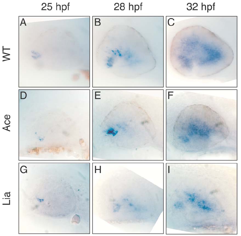
Fig. S3 Expression of Ath5 in zebrafish wt and Fgf mutant embryos. The temporal and spatial expression of Ath5 was examined in wild type (A,B,C), Ace/Fgf8 (D,E,F) and Lia/Fgf3 (G,H,I) mutant embryos. No significant difference was observed at 25 hpf and 28 hpf (A, D, G and B, E, H, respectively). However by 32 hpf a slight decrease in Ath5 expression was observed in Ace and Lia mutants (compare C with F and I). This could be due to a secondary developmental defect of these embryos or alternatively it would support a partially non-redundant role of Fgf8 and Fgf3 for the correct progression of Ath5 expression and retinal ganglion cell differentiation.
Reprinted from Developmental Cell, 8(4), Martinez-Morales, J.R., Del Bene, F., Nica, G., Hammerschmidt, M., Bovolenta, P., and Wittbrodt, J., Differentiation of the vertebrate retina is coordinated by an FGF signaling center, 565-574, Copyright (2005) with permission from Elsevier. Full text @ Dev. Cell