
Image
|
Figure Caption
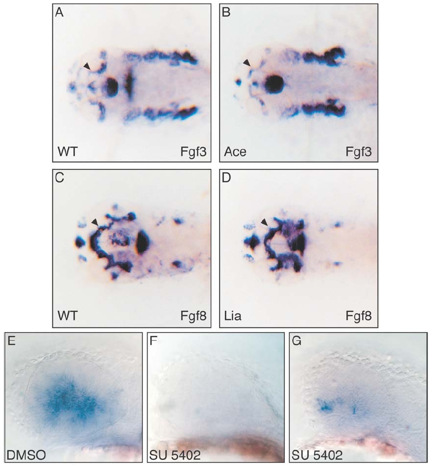
Fig. S2 Expression of Fgf3 and Fgf8 in zebrafish Fgf mutant embryos. The expression of Fgf3 and Fgf8 was analyzed in Ace/Fgf8 and Lia/Fgf3 mutant embryos, respectively. No difference was observed in the expression of these genes in the optic stalk region, indicated by arrowheads (compare A with B, and C with D). This excludes any feedback cross regulation or genetic hierarchy between these two genes in this domain.
Inhibition of Fgf signalling prevents Ath5 expression at 32 hpf. Drug treatment with the Fgf inhibitor SU5402 abolishes or strongly reduces Ath5 expression (compare E with F and G). Embryos where treated from 23 to 32 hpf. The mock DMSO treated embryos showed always an extended Ath5 expression (E, 12 out of 12) while the SU5402 treatment either prevents Ath5 expression (F, 5 out of 11) or results in a strongly reduced expression (G, 6 out of 11).
Figure Data
Acknowledgments
This image is the copyrighted work of the attributed author or publisher, and
ZFIN has permission only to display this image to its users.
Additional permissions should be obtained from the applicable author or publisher of the image.
Reprinted from Developmental Cell, 8(4), Martinez-Morales, J.R., Del Bene, F., Nica, G., Hammerschmidt, M., Bovolenta, P., and Wittbrodt, J., Differentiation of the vertebrate retina is coordinated by an FGF signaling center, 565-574, Copyright (2005) with permission from Elsevier. Full text @ Dev. Cell