
|
Fig. S1
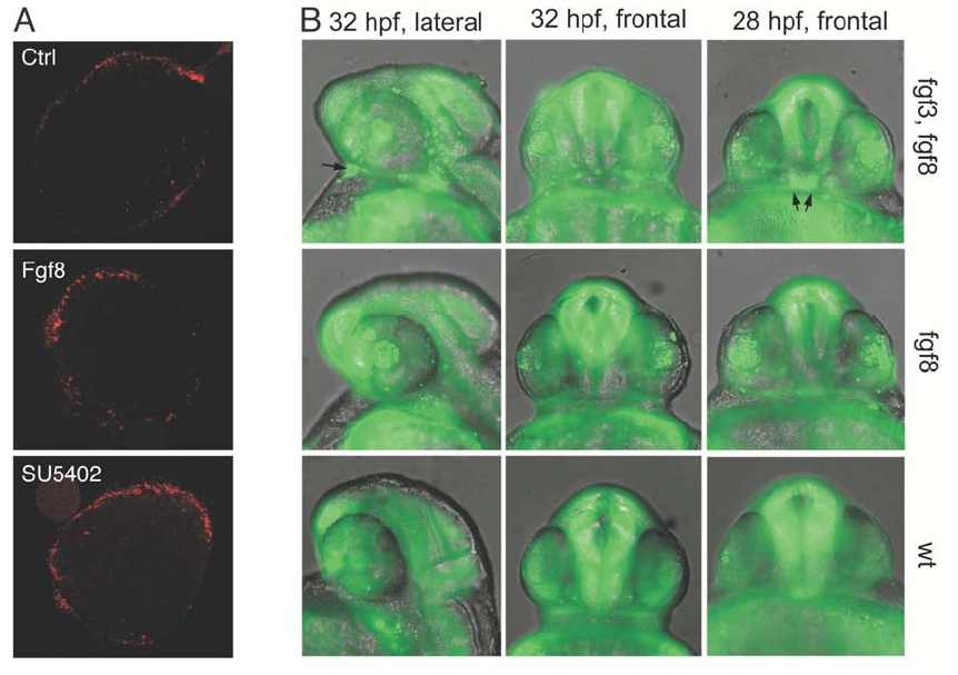
(A) Cell death was examined in control explants and explants exposed to FGF8 or SU5402 by propidium iodide staining. Note that the amount of cell death is minimal and basically limited to the edges in all explants under the experimental conditions employed.
(B) Analysis of apoptosis in zebrafish Fgf mutants. Increased apoptosis was observed in the retina and lens of Lia/Fgf3 and Ace/Fgf8 double mutants compared to the control. On the contrary Ace/Fgf8 single mutants showed increased apoptosis in the lens but not in the retina. There is no obvious increase of apoptosis in the optic stalk region.
Reprinted from Developmental Cell, 8(4), Martinez-Morales, J.R., Del Bene, F., Nica, G., Hammerschmidt, M., Bovolenta, P., and Wittbrodt, J., Differentiation of the vertebrate retina is coordinated by an FGF signaling center, 565-574, Copyright (2005) with permission from Elsevier. Full text @ Dev. Cell