
Image
|
Figure Caption
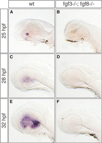
Fig. 6 Fgf3 and Fgf8 Cooperate in Initiating Retina Differentiation
(A–F) Lateral views of (B, D, and F) Fgf3 and Fgf8 double mutant embryos and (A, C, and E) wild-type siblings at 25 hpf, 28 hpf, and 32 hpf hybridized with an ath5-specific probe. In contrast to wild-type, ath5 transcripts are absent in mutant embryos (compare [B], [D], and [F] to [A], [C], and [E], respectively). Dashed lines demarcate the outlining of the eye.
Figure Data
Acknowledgments
This image is the copyrighted work of the attributed author or publisher, and
ZFIN has permission only to display this image to its users.
Additional permissions should be obtained from the applicable author or publisher of the image.
Reprinted from Developmental Cell, 8(4), Martinez-Morales, J.R., Del Bene, F., Nica, G., Hammerschmidt, M., Bovolenta, P., and Wittbrodt, J., Differentiation of the vertebrate retina is coordinated by an FGF signaling center, 565-574, Copyright (2005) with permission from Elsevier. Full text @ Dev. Cell